HVF Test in Andheri West, Mumbai — Visual Field Test Cost, Procedure & How to Read Your Report, by a Specialist
Written & medically reviewed by Dr. Jignesh Gala, Vitreoretinal & Cataract Surgeon (FRCS Glasgow), Founder & Director — Crystal Clear Eye Clinic (Andheri West) and Topax Eye Hospital (Vile Parle East), Mumbai. Prices & facts verified: 24 July 2026.
⚡ Quick answer: An HVF test (Humphrey Visual Field test, also called perimetry or a “field test”) maps everything your eye can see — centre and side vision — by asking you to press a button whenever you notice a tiny light. It is the gold-standard functional test for glaucoma, and also detects field loss from stroke, pituitary tumours, ptosis and drug toxicity. Searching for an HVF test near me or the visual field test cost in Mumbai? Expect ₹750–1,600 per eye at clinics (CGHS reference: ₹800 both eyes). At Crystal Clear Eye Clinic, Andheri West, your field is tested on a ZEISS Humphrey Field Analyzer and every report — MD, PSD, VFI, GHT — is read and explained by the doctor himself. Call 077188 85245.
1. What Is an HVF (Visual Field) Test?
HVF = Humphrey Visual Field — the world’s most trusted “field of vision” test, performed on the ZEISS Humphrey Field Analyzer (the machine is the reason for the name). In simple words (HVF test kya hota hai?): your vision is not just the sharp centre you read with — it is also everything you see “from the corner of your eye.” The HVF test maps that entire island of vision, point by point, by flashing dim lights in a white bowl while you press a button for each one you see. Every point you miss tells a story — and in glaucoma, that story begins years before you notice anything wrong.
Doctors also call it automated perimetry — and patients call it the “button test,” the “light test,” or simply the eye field test / field test for eyes. Whatever the name, it answers one question no other test can: how much of your world can you actually see? That is why the visual field test remains the medico-legal and scientific gold standard for diagnosing glaucoma, judging its severity, and — most importantly — proving whether it is getting worse. If you searched for a visual field test near me, a perimetry test near me, or an HVF test in Andheri West, this page covers everything: cost, procedure, and the report-reading knowledge no other Mumbai clinic publishes.
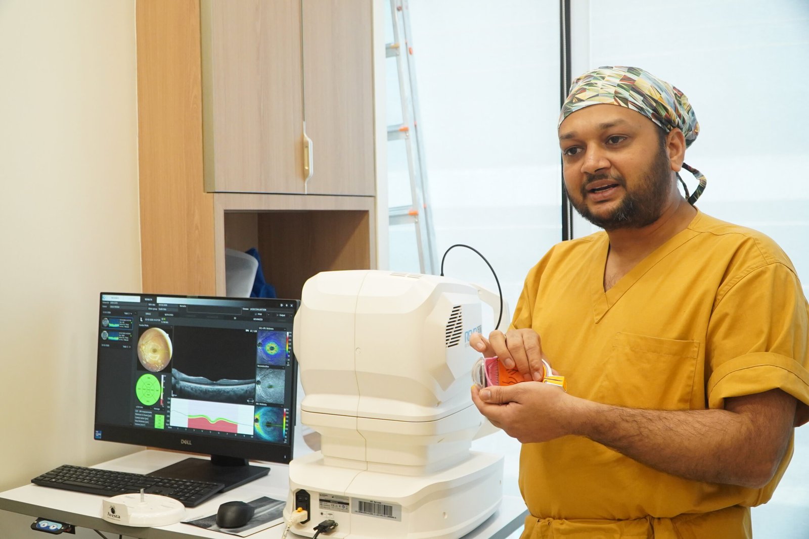
A crucial point most clinics never explain: a field test is a functional test — it measures what your eye does, not just what it looks like. Eye pressure is a number; OCT is a photograph; but the HVF is your actual seeing ability, quantified. Glaucoma treatment has one goal — protecting your field of vision — so the field test is the scoreboard of that entire effort. Dr. Jignesh Gala, glaucoma and retina specialist in Andheri West, personally correlates every field with your eye pressure, OCT and examination before any treatment decision.
2. Test Types: 24-2, 24-2C, 10-2, SITA Strategies & Esterman
When a lab says “field test,” ask which programme and which strategy — the choice changes what the test can find:
| Programme | What It Maps | Used For |
|---|---|---|
| 24-2 (most common) | 54 points across the central 24° — where glaucoma strikes first | Standard glaucoma diagnosis & follow-up; most neurological screening |
| 24-2C (newer) | 24-2 pattern plus 10 extra central points | Catches early central/macular field loss that classic 24-2 misses — Dr. Gala’s preferred glaucoma programme |
| 10-2 | 68 points packed into the central 10° | Advanced glaucoma threatening fixation, macular disease, hydroxychloroquine toxicity screening |
| 30-2 | Wider 30° field, 76 points | Neurological fields (stroke, pituitary), broader screening — longer test |
| Esterman (binocular) | Whole binocular field, both eyes open | Driving licence / fitness certification — the statutory standard |
| Ptosis protocol | Superior field with eyelid taped vs untaped | Documenting functional impairment for ptosis surgery insurance approval |
And the strategies (the algorithm that runs the test): SITA Standard is the classic validated algorithm; SITA Fast is quicker for screening; SITA Faster — the current generation — cuts test time to ~2–4 minutes per eye without losing accuracy, which means less fatigue and fewer reliability errors for elderly patients. Faster, smarter testing is one reason the machine generation matters: an old perimeter running a 15-minute Swedish test exhausts patients into unreliable results. At Dr. Gala’s centres, strategies are chosen per patient — a 35-year-old screening patient and an 80-year-old with advanced glaucoma do not get the same test.
3. HVF vs OCT — Function vs Structure (Which One Do You Need?)
The most common confusion in glaucoma care: “Doctor, my OCT was done last month — why do I need a field test?” Because they measure different things, and glaucoma care needs both:
| HVF (Visual Field / Perimetry) | OCT | |
|---|---|---|
| Measures | Function — what you actually see (sensitivity at each point) | Structure — anatomy (nerve fibre layer thickness) |
| Detects glaucoma | When nerve damage is enough to affect vision | Often earlier — structural thinning can precede field loss |
| Tracks worsening | VFI trend (%/year) — the legal & clinical severity scoreboard | RNFL trend — useful until “floor effect” in advanced disease |
| Also detects | Stroke, pituitary tumours, ptosis impact, drug toxicity | Macular diseases, edema, holes, AMD |
| Test experience | Active — you press a button for 3–7 min/eye | Passive — machine scans in seconds |
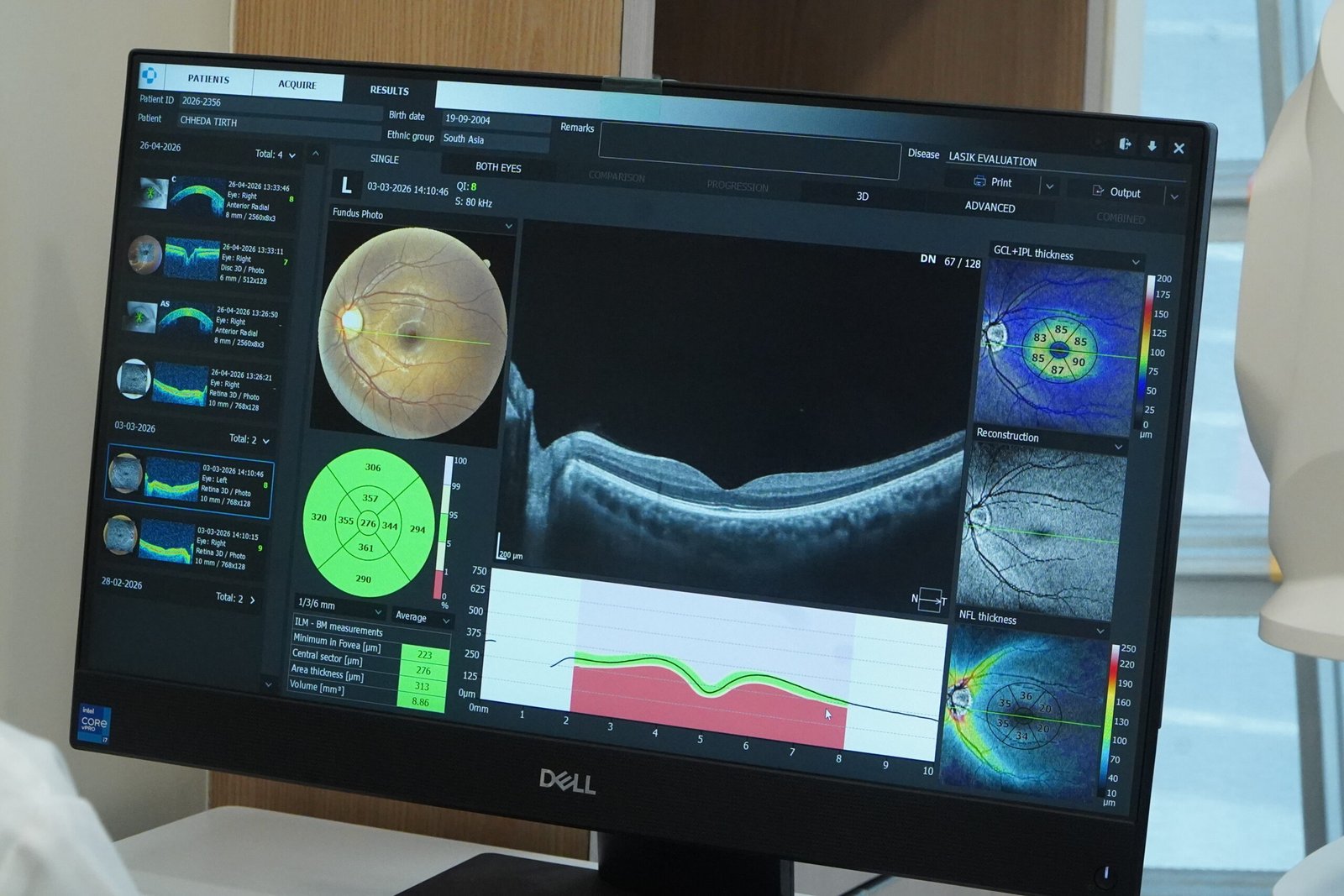
The structure–function relationship is one of the most researched areas in glaucoma (PubMed evidence): sometimes the OCT changes first, sometimes the field does — which is why international guidelines require both for diagnosis and follow-up. Rule of thumb: OCT for early detection, HVF for severity and progression, both together for decisions. Any clinic following only one of the two is flying with one wing.
4. How to Read Your HVF Report — MD, PSD, VFI, GHT Explained
No clinic in Mumbai publishes this section. Patients deserve to understand their own printout — so here it is.
Your single-page ZEISS printout has four zones. Spend two minutes with this table and you will read your report better than most patients anywhere:
| Report Term | Full Form | What It Tells You (in plain words) |
|---|---|---|
| Reliability indices (fixation losses, false +ve, false −ve) | — | Can this test be trusted at all? High fixation losses = you weren’t looking at the centre; high false positives = trigger-happy button pressing. An unreliable test is repeated, not interpreted. |
| GHT | Glaucoma Hemifield Test | The machine’s built-in glaucoma suspicion: “Within Normal Limits” (good), “Borderline”, or “Outside Normal Limits” (abnormal). |
| MD | Mean Deviation | Your overall depression vs a healthy person your age. 0 to −2 dB ≈ normal; −2 to −6 early; −6 to −12 moderate; worse than −12 advanced. MD is the number Dr. Gala tracks at every visit. |
| PSD | Pattern Standard Deviation | How uneven your field is — the higher the PSD, the more localized the damage (typical of glaucoma). |
| VFI | Visual Field Index | Your field as a simple percentage (100% = perfect). VFI 95%+ early; below 80% significant; below 60% severe. The easiest number for patients to follow — and the one used in progression analysis. |
| Grayscale map | — | The grey picture: dark patches = areas of poor sensitivity (scotoma). Dramatic to look at, but doctors actually read the numbers beside it. |
| Total & Pattern deviation plots | — | Point-by-point comparison with normal. The pattern deviation plot removes overall haze (like cataract) to reveal true localized nerve damage — the plot glaucoma specialists stare at longest. |
Why reliability matters first: a famous perimetry truth — the first field test of your life is often “worse” than reality, simply because the test has a learning effect (evidence). That is why Dr. Gala never changes treatment on a single new-worsening field: he confirms with a repeat test. Reliability indices decide whether a report is even worth reading (reliability evidence) — and this single discipline prevents countless unnecessary medicine changes across Mumbai.
5. 8 Conditions Detected by Visual Field Testing
- Glaucoma — the #1 reason for a glaucoma field test. Field loss patterns (arcuate scotoma, nasal step) are diagnostic, and serial fields judge control (detection evidence). Every glaucoma patient needs baseline + lifelong periodic fields.
- Ocular hypertension & glaucoma suspects — high eye pressure with a normal field is watched; the first field defect converts diagnosis to glaucoma and starts treatment (OHTS evidence).
- Stroke & brain lesions — a homonymous hemianopia (same-side half-field loss in both eyes) points behind the eyes, to the brain (neuro evidence). The field test often detects what the patient only felt as “bumping into things.”
- Pituitary tumours — classically cause bitemporal hemianopia (losing the outer halves in both eyes). Many pituitary adenomas are first suspected on a field chart, not an MRI.
- Ptosis (droopy eyelid) — superior field defects document functional vision loss, the evidence insurers demand before approving ptosis surgery (ptosis field evidence).
- Hydroxychloroquine (HCQ) toxicity — patients on long-term HCQ (arthritis, lupus, skin disease) need annual 10-2 fields to catch retinal toxicity early (screening evidence).
- Driving fitness & occupational certification — the binocular Esterman field is the standard for licence/disability assessment; field loss impairs driving safety measurably (driving evidence).
- Central/macular field loss — the 10-2 programme catches early central damage that standard 24-2 testing misses (10-2 evidence) — vital in advanced glaucoma where every central point matters.
6. Who Needs an HVF & How Often
| You Are… | Recommended Field Test | How Often |
|---|---|---|
| A diagnosed glaucoma patient | 24-2 or 24-2C (SITA Faster); 10-2 if advanced | Every 6–12 months; more often if progressing or after treatment change |
| Glaucoma suspect / ocular hypertensive / family history | 24-2 baseline | Every 6–12 months; 2–3 baselines in the first 2 years to establish your true starting point |
| 40+ with diabetes, high myopia, or steroid use | Screening field as advised | Yearly with your glaucoma workup |
| On long-term hydroxychloroquine | 10-2 (+ OCT) | Baseline, then annually after 5 years (earlier if risk factors) |
| Droopy eyelid bothering vision | Ptosis protocol (taped/untaped) | Once, for surgical documentation |
| Post-stroke / brain tumour follow-up | 30-2 or as the neurologist requests | Per neurology plan |
| Driving licence / fitness / disability certificate | Binocular Esterman | As required by authority |
7. The Test Procedure — Step by Step (HVF test mein kya hota hai)
Patients ask visual field test kaise hota hai (aankhon ka field test) — here is the honest walkthrough:
- No preparation (0 min): No fasting, no injections, no dilating drops for the field test itself. Bring your current glasses — the test is done with your correct prescription in the machine’s lens holder.
- Setup (2 min): One eye is gently patched. You sit at the white bowl of the ZEISS Humphrey Field Analyzer, chin on the rest, staring at a central yellow light.
- The test (3–7 min per eye with SITA Faster): Small lights flash at different spots and brightnesses. Press the button every time you notice one — even if barely. You are not supposed to see every light; the machine deliberately shows some you cannot see. That is how it finds your threshold.
- Mistakes are normal: pressed by accident? missed one? The machine measures false responses and fixation automatically — these become the reliability indices. You cannot “fail” by being human.
- Second eye, then done: total visit usually 20–30 minutes. No after-effects — drive yourself home, back to work the same day.
- Report & explanation: the printout is generated instantly. At Dr. Gala’s centres, the doctor walks you through MD, PSD, VFI and the maps on the report itself — and stores every field digitally so your next visit’s test is compared point-by-point.
HVF test ke nuksan (disadvantages)? Honestly: it needs your attention for a few minutes, and the first-ever test can feel confusing — that’s why we explain before we start, and why SITA Faster’s shorter test suits elderly patients. No pain, no drops, no radiation, no side effects.
8. Glaucoma Progression Tracking — Guided Progression Analysis (GPA)
Diagnosing glaucoma is the beginning. The real question for the rest of life: is it getting worse, and how fast? This is where the ZEISS Guided Progression Analysis (GPA) changes everything — and why fields must always be done on the same machine, at the same centre:
- Baseline fields: your first 2–3 reliable tests become the reference. (One baseline is never enough — see the learning effect above.)
- Every follow-up field is auto-compared to baseline: GPA flags points that have worsened beyond normal test-to-test variability — “Possible Progression” or “Likely Progression.”
- VFI trend analysis plots your Visual Field Index over years and calculates your rate of change in %/year — the single most important number in glaucoma. A patient losing 2%/year needs escalation now; one stable at 0.1%/year is safely coasting (GPA evidence, progression evidence).
- Why it must be the same machine: different machines (Humphrey vs Octopus vs FDP screening units) speak different languages — their results cannot be trended together. This is why Dr. Gala’s patients keep their field record at his centres: your 2026 field talks to your 2031 field.

The honest truth about field testing in Mumbai: many centres do a field test only when “the doctor asks for it,” print it, file it — and never trend it. Glaucoma is not managed by single reports; it is managed by slopes. At Crystal Clear Eye Clinic and Topax Eye Hospital, every new field is plotted against your history, and treatment decisions follow the trend, not the day. With SITA Faster shortening tests (SITA Faster evidence) and modern algorithms improving reliability (SITA evidence), there is no excuse for managing glaucoma 2005-style in 2026.
9. HVF Test Cost in Mumbai & Andheri West (2026 Price Table)
Transparent, date-stamped pricing (verified 24 July 2026) — because only one Mumbai clinic in the entire city publishes its visual field price, and patients deserve better:
| Test | Typical Mumbai Range | What Others Quote | At Dr. Gala’s Centres |
|---|---|---|---|
| HVF / visual field test (per eye) | ₹750–1,600 | ₹800/eye (a Mumbai chain); ₹750–950/eye (aggregator clinics); up to ₹3,000 (premium) | Within the normal city band — interpreted by the doctor, trended against your history |
| HVF both eyes | ₹1,200–3,000 | ₹1,600 both (chain price); CGHS reference rate: ₹800 both eyes (metro, NABH) | Honest, quoted upfront on call — no surprises |
| Esterman (driving/binocular) | ₹800–1,500 | Rarely quoted anywhere | Available with proper certification format |
| Glaucoma workup package (field + OCT RNFL + pressure + pachymetry) | ₹2,500–5,000 | Chains bundle ~15% off, “under ₹5,000” | Complete specialist workup — every number correlated by Dr. Gala himself |
Honest guidance: the HVF test price across Mumbai varies within a fairly narrow band — a field test is not where clinics make their margin. What you are really choosing is who reads it and whether it gets trended. A ₹500 field filed away unread is money wasted; a properly interpreted, digitally stored, GPA-trended field is an investment that guides every glaucoma decision for years. Ask our team today’s exact price on 077188 85245 — you will get a number, not a runaround. And if you are comparing the visual field test cost in Mumbai or the HVF test cost in Andheri West, compare the interpretation that comes with it, not just the rupee figure.
10. Insurance, Cashless & CGHS
- Standalone OPD field tests are usually outside standard mediclaim OPD cover — true everywhere in Mumbai. OPD-cover policies and corporate health cards increasingly reimburse diagnostics; we provide GST invoices and machine-signed printouts that claim offices accept.
- When the field test is part of a covered procedure (glaucoma surgery package, ptosis surgery documentation), it is typically bundled into the claim. Here Dr. Gala’s dual setup helps: direct cashless is done through Topax Eye Care (Vile Parle East), and third-party/TPA cashless is done through Crystal Clear Eye Clinic (Andheri West).
- CGHS / reimbursement patients: rates stay within standard reference bands (CGHS reference ₹800 both eyes), with documentation in the exact format reimbursement desks expect.
- Ptosis surgery patients: we perform the taped/untaped superior field documentation insurers specifically demand before approving functional ptosis correction.
11. Why Patients Choose Dr. Jignesh Gala for Visual Field Testing

Dr. Jignesh Gala is an FRCS (Glasgow)-trained eye surgeon — Founder & Director of both Crystal Clear Eye Clinic (Andheri West) and Topax Eye Hospital (Vile Parle East). For glaucoma patients searching for a glaucoma specialist Andheri West trusts — the best glaucoma specialist in Andheri West or an HVF test in Andheri West, his centres offer something diagnostic labs cannot: the test, the interpretation, and the treatment — all under one roof, by one accountable doctor.
✅ ZEISS Humphrey Field Analyzer — the global gold-standard perimeter, running modern SITA strategies including SITA Faster for shorter, more reliable tests in elderly patients.
✅ Every report read by the doctor himself — MD, PSD, VFI, GHT and the deviation plots explained on the printout, in Hindi, Marathi, Gujarati or English. Never a technician handover, never “report is normal, don’t worry.”
✅ Progression-trended records: your fields are stored and compared year-on-year with Guided Progression Analysis — the discipline that actually prevents glaucoma blindness.
✅ Fully aware of the latest research — unlike old practitioners who stopped reading journals: 24-2C central-point strategies, structure–function correlation, home/VR perimetry developments — 400+ verified references below are the evidence base he follows.
✅ Honest retest policy: a first-time or unreliable field is repeated, not acted upon. No treatment escalations on single suspicious reports.
12. Locations — HVF Test in Andheri West & Vile Parle East
1) Crystal Clear Eye Clinic — Andheri West
📍 Address: Laram Centre CHS, A1-202, Swami Vivekanand Road, above Sunil Jewellers, near NADCO Shopping Centre, Railway Colony, Andheri West, Mumbai 400058
📞 Phone: 077188 85245 • 🕘 Open daily, closes 9 pm
💳 Third-party / TPA cashless is done through Crystal Clear Eye Clinic.
Minutes from Andheri station, Lokhandwala, Versova, Yari Road, Juhu, Oshiwara, DN Nagar, Seven Bungalows, Azad Nagar, Veera Desai Road, Jogeshwari West, Vile Parle West and Amboli. Patients searching HVF test near me, visual field test in Andheri West or perimetry test near me from Bandra, Khar, Santacruz, Goregaon, Malad and Kandivali reach us in 20–30 minutes.

2) Topax Eye Hospital — Vile Parle East
📍 Vile Parle East, Mumbai (full address on drjigneshgalaeye.com) • 📞 077188 85245
💳 Direct cashless is done through Topax Eye Care.
For an HVF test in Vile Parle East / visual field test in Vile Parle — convenient for Santacruz East, Khar, Bandra East, Kalina, Vakola, Juhu and the airport corridor. As a full hospital, Topax handles everything your field test may lead to — medicines, lasers (SLT), glaucoma surgery — under Dr. Gala himself.
Distance is not a barrier: glaucoma patients travel from Thane, Navi Mumbai, Borivali and Dadar for trended field testing. A visual field record is a lifetime asset — keep it with a specialist who will still be reading your slopes a decade from now.
13. हिंदी • मराठी • ગુજરાતી — Patient Summaries
🇮🇳 हिंदी सारांश — विज़ुअल फील्ड टेस्ट (एचवीएफ)
विज़ुअल फील्ड टेस्ट क्या है? यह आँख के दृष्टि क्षेत्र (सामने + किनारों की नज़र) का नक्शा बनाने वाली बिल्कुल बेदर्द जांच है — मशीन में छोटी रोशनी दिखने पर बटन दबाना होता है। यह ग्लूकोमा की जांच (ग्लूकोमा (काला मोतिया) की जांच) का सबसे भरोसेमंद तरीका है और ब्रेन ट्यूमर, स्ट्रोक तथा पलक झुकने से होने वाली दृष्टि हानि भी पकड़ता है। मुंबई में कीमत ₹750–1,600 प्रति आँख। ग्लूकोमा विशेषज्ञ अंधेरी वेस्ट में — डॉ. जिग्नेश गाला ZEISS Humphrey मशीन पर टेस्ट कराके MD, PSD, VFI समेत पूरी रिपोर्ट खुद आपकी भाषा में समझाते हैं। अपॉइंटमेंट: 077188 85245।
🚩 मराठी सारांश — व्हिज्युअल फील्ड टेस्ट (एचव्हीएफ)
व्हिज्युअल फील्ड टेस्ट म्हणजे काय? डोळ्याच्या दृष्टिक्षेत्राचा (समोरची व बाजूची दृष्टी) नकाशा तयार करणारी पूर्ण वेदनारहित तपासणी — छोटा दिवा दिसल्यावर बटण दाबायचे. ग्लुकोमाची (काळा मोतिया) तपासणी होण्यासाठी ही सर्वांत महत्त्वाची चाचणी; स्ट्रोक, पिट्युटरी ट्यूमर व डोळकणे सारख्या समस्याही यातून धरल्या जातात. मुंबईत किंमत ₹७५०–१,६०० प्रति डोळा. ग्लुकोमा तज्ज्ञ अंधेरी वेस्ट येथे — डॉ. जिग्नेश गाला ZEISS Humphrey मशीनवर तपासणी करून अहवाल स्वतः मराठीत समजावतात. भेटीसाठी: 077188 85245.
🟡 ગુજરાતી સારાંશ — વિઝ્યુઅલ ફિલ્ડ ટેસ્ટ (HVF)
વિઝ્યુઅલ ફિલ્ડ ટેસ્ટ શું છે? આંખના દૃષ્ટિક્ષેત્રનો (સામેની + બાજુની નજર) નકશો બનાવતી સંપૂર્ણ પીડારહિત તપાસ — નાનો દીવો દેખાય ત્યારે બટન દબાવવાનું. ગ્લૂકોમા (કાળા મોતિયા)ની તપાસ માટેની સૌથી અગત્યની કસોટી; સ્ટ્રોક, પિટ્યુટરી ટ્યૂમર અને પલક ઢળવાથી થતી નુકસાન પણ પકડે છે. મુંબઈમાં કિંમત ₹૭૫૦–૧,૬૦૦ દરેક આંખ. ગ્લૂકોમા સ્પેશિયાલિસ્ટ અંધેરી વેસ્ટમાં — ડૉ. જિગ્નેશ ગાલા ZEISS Humphrey મશીન પર ટેસ્ટ કરાવીને રિપોર્ટ જાતે ગુજરાતીમાં સમજાવે છે. એપોઈન્ટમેન્ટ: 077188 85245.
14. ❓ 12 Frequently Asked Questions (English + Hinglish)
1. HVF test dard hota hai kya? (Is the visual field test painful?)
Bilkul nahi. Koi injection nahi, koi drops nahi, machine aapki aankh ko touch bhi nahi karti. Aap bas white bowl ki taraf dekhte hain aur chhoti light dikhe toh button dabate hain — 3 to 7 minutes per eye. The only “effort” is paying attention. No pain, no side effects, no recovery time.
2. HVF test kitne minute lagte hain? (How long does it take?)
With the modern SITA Faster strategy: about 3–7 minutes per eye. Older machines/strategies take 10–15 minutes per eye. Including setup for both eyes, plan for a 20–30 minute visit. Report is printed instantly.
3. HVF test cost in Andheri West – kitna kharcha aata hai?
Mumbai range: ₹750–1,600 per eye (chains quote ₹800/eye or ₹1,600 both; CGHS reference ₹800 both eyes). At Crystal Clear Eye Clinic you pay within the normal city band — and the doctor himself reads and trends your report. Visual field test ki kimat confirm karne ke liye call 077188 85245.
4. What is the difference between HVF and OCT?
HVF measures function — what your eye actually sees, point by point. OCT measures structure — the anatomy/thickness of the nerve layer. Glaucoma needs both: OCT catches early structural damage; HVF proves functional loss and tracks your vision over the years. Think: OCT is the photograph of the wiring; HVF is whether the lights still turn on.
5. HVF report mein MD, PSD, VFI, GHT ka kya matlab hai?
GHT = machine’s glaucoma suspicion (Within/Outside Normal Limits). MD = overall vision depression vs normal (0 to −2 dB normal; worse than −12 advanced). PSD = how uneven/patchy the damage is. VFI = your field as a percentage (100% = perfect) — the easiest number to follow. Reliability indices on top tell whether the test itself can be trusted. Full patient guide in Section 4 above — the only one published by any Mumbai clinic.
6. Visual field test kyun zaroori hai glaucoma mein?
Kyunke glaucoma ka ilaaj sirf ek maqsad ke liye hai — aapki nazariye ka kshetra (field of vision) bachana. Eye pressure toh sirf ek number hai; asli result ye hai ki aapki field stable hai ya ghatti ja rahi hai. Bina field test ke glaucoma treatment andheron mein teer chalane jaisa hai — isliye har 6–12 mahine mein field repeat hoti hai, aur har field aapki purani fields se compare hoti hai.
7. Field test mein kitne lights dikhani chahiye? Bahut miss hue toh?
Relax — you are supposed to miss many lights. The machine deliberately shows dimmer and dimmer lights to find your threshold; “missing” is built into the design. It also plants fake checks (lights you should see easily, and moments with no light at all) to measure false responses. Your job is only to be honest: press when you see, don’t press when you don’t. The computer handles the rest.
8. How often should the visual field test be repeated in glaucoma?
Typically every 6–12 months for stable patients; every 3–6 months if progression is suspected, after any treatment change, or in advanced disease. Newly diagnosed patients need 2–3 baseline fields in the first two years. Always on the same machine, at the same centre — otherwise the trend comparison (GPA) is impossible.
9. Can a visual field test detect brain tumor or stroke?
Yes — this surprises most patients. The pattern of field loss localizes the problem: bitemporal hemianopia (outer halves missing in both eyes) classically signals a pituitary tumour pressing the optic chiasm; homonymous hemianopia (same side missing in both eyes) points to the brain — stroke, tumour or injury behind the eye. Many patients are referred for MRI on the basis of a field chart alone.
10. HVF test kahan karwaye Andheri West mein?
Crystal Clear Eye Clinic — Laram Centre CHS, A1-202, S.V. Road, above Sunil Jewellers, near NADCO Shopping Centre, Andheri West 400058. Or Topax Eye Hospital, Vile Parle East. Same phone for both: 077188 85245. Open daily, closes 9 pm. Same-day appointments usually available; report in hand immediately.
11. Is a visual field test needed for driving licence or ptosis surgery?
Yes to both, when asked. For driving/fitness certification, the binocular Esterman field test is the accepted standard. For ptosis (droopy eyelid) surgery, insurers demand a taped-vs-untaped superior field proving the lid blocks functional vision — we perform exactly that documentation. Bring the authority’s format if you have one.
12. Test ke dauran button galat dab gaya toh report kharab ho jayegi kya?
Nahi — a few wrong presses are completely normal and expected. The machine continuously measures false positives (pressing with no light), false negatives (not pressing bright lights) and fixation losses (eye wandering). Only if these cross limits is the test declared “unreliable” — and then we simply repeat it, often with the shorter SITA Faster strategy. Ek galat button se report kharab nahi hoti.
📖 Related Reading by Dr. Jignesh Gala
Keep learning from the same specialist who reads your field tests:
- OCT Eye Test in Andheri West, Mumbai — cost, procedure & report explained
- Intravitreal Injection in Andheri West, Mumbai — the complete guide
- Anti-VEGF Injection Cost in Mumbai — 2026 honest price guide
- Retina Laser Treatment in Andheri West / Mumbai
- Best Retina Specialist in Mumbai — Dr. Jignesh Gala
- Best Eye Specialist in Andheri, Mumbai
- Retina Treatment in Andheri
- Retina Treatment in Mumbai — Advanced Retinal Care
- Retina Surgery in Mumbai — Your Complete Guide to Saving Your Sight
- Cashless Eye Surgery in Mumbai
- Surgical Videos by Dr. Jignesh Gala
- Inside My Six Years as a BMJ Case Reports Peer Reviewer
- Diabetic Retinopathy in Andheri
- Diabetic Eye Care in Mumbai
- Diabetic Macular Edema (DME) Treatment in Mumbai
- Macular Edema Treatment in Andheri West
- ARMD Treatment in Andheri West, Mumbai
- Vitreoretinal Treatment in Andheri
- Best Retina Specialist in Mumbai
- Best Vitrectomy Surgery in Mumbai — Expert Retinal Care
- Cashless Eye Surgery in Mumbai
- Sudden Floaters? Retina Check-up in Mumbai
- What Is an Intravitreal Eye Injection? Explained by Dr. Jignesh Gala
Glaucoma steals vision silently. A field test is how you catch it.
Whether you searched for an HVF test near me, a visual field test in Mumbai, a perimetry test near me, or checked the HVF test cost in Andheri West — you have found transparent pricing, a ZEISS Humphrey Field Analyzer, and a specialist who reads every single report himself. One 20-minute visit today protects the vision you will need for the next 20 years.
📍 Andheri West: Laram Centre CHS, A1-202, S.V. Road, above Sunil Jewellers, near NADCO Shopping Centre, Mumbai 400058
📍 Vile Parle East: Topax Eye Hospital • Open daily, closes 9 pm
Dr. Jignesh Gala, FRCS (Glasgow) — Eye Surgeon • drjigneshgalaeye.com • crystalcleareye.in
Medical disclaimer: This page is educational and does not replace a personal consultation. Prices are indicative, verified on 24 July 2026, and may change; please confirm at booking. Every eye is different — glaucoma decisions are made only after examination and field correlation by Dr. Jignesh Gala.
15. Research & Evidence Library — 400+ Verified References
Every link below was verified live in July 2026 via the official NCBI E-utilities, ClinicalTrials.gov v2 API, CrossRef, Europe PMC and publisher pages. 508 links across PubMed, PubMed Central-indexed journals, Google Scholar, the Cochrane Library, Europe PMC, ClinicalTrials.gov, Ophthalmology, Ophthalmology Glaucoma, the American Journal of Ophthalmology, the Journal of Glaucoma, IOVS, TVST, the British Journal of Ophthalmology, JAMA Ophthalmology, Eye (Nature), the Indian Journal of Ophthalmology, Acta Ophthalmologica, Graefe’s Archive, the European Glaucoma Society and ZEISS Humphrey Field Analyzer resources — the same evidence Dr. Gala reads. Click any title to verify the science yourself.
🔬 Visual Field & Perimetry Clinical Evidence — PubMed (393 verified)
Humphrey Visual Field & Progression
- Visual field progression in patients with severe glaucoma — Graefe’s archive for clinical and experimental ophthalmology = Albrecht von Graefes Archiv fur klinische und experimentelle Ophthalmologie, 2021
- Visual Field Progression in Glaucoma During COVID-19 — Journal of glaucoma, 2025
- Early Manifest Glaucoma Trial: design and baseline data — Ophthalmology, 1999
- Risk factors for visual field progression in newly diagnosed exfoliation glaucoma patients in Sweden — Scientific reports, 2022
- Predicting Visual Field Progression by Optical Coherence Tomography Angiography and Pattern Electroretinography in Glaucoma — Journal of glaucoma, 2022
- Reduction of intraocular pressure and glaucoma progression: results from the Early Manifest Glaucoma Trial — Archives of ophthalmology (Chicago, Ill. : 1960), 2002
- Visual Field Progression in Glaucoma Patients With Delayed Follow-Up — American journal of ophthalmology, 2026
- Visual Subfield Progression in Glaucoma Subtypes — Journal of ophthalmology, 2018
- Comparison of visual field progression in new-diagnosed primary open-angle and exfoliation glaucoma patients in Sweden — BMC ophthalmology, 2020
- Distribution and Progression of Visual Field Defects With Binocular Vision in Glaucoma — Journal of glaucoma, 2018
- To identify risk factors for central visual field progression in moderate to advanced glaucoma — Indian journal of ophthalmology, 2024
- Visual field progression in glaucoma: what is the specificity of the Guided Progression Analysis? — Ophthalmology, 2014
- Risk factors for visual field progression in treated glaucoma — Archives of ophthalmology (Chicago, Ill. : 1960), 2011
- To compare central visual field progression using mean deviation and pointwise linear regression analysis — Indian journal of ophthalmology, 2023
- Relationship between visual field progression and baseline refraction in primary open-angle glaucoma — Clinical ophthalmology (Auckland, N.Z.), 2016
- Progression of visual field in patients with primary open-angle glaucoma – ProgF study 1 — Acta ophthalmologica, 2015
- Improving the prediction of visual field progression in glaucoma using spatial processing — Ophthalmology, 1997
- Frontloading visual field tests detect earlier mean deviation progression when applied to real-world-derived early-stage glaucoma data — Ophthalmic & physiological optics : the journal of the British College of Ophthalmic Opticians (Optometrists), 2024
- Former smoking as a risk factor for visual field progression in exfoliation glaucoma patients in Sweden — European journal of ophthalmology, 2024
- Interobserver agreement on visual field progression in glaucoma: a comparison of methods — The British journal of ophthalmology, 2003
- Glaucoma Progression Analysis software compared with expert consensus opinion in the detection of visual field progression in glaucoma — Ophthalmology, 2012
- Changes in Axial Length and Progression of Visual Field Damage in Glaucoma — Investigative ophthalmology & visual science, 2018
- Survey on electronic visual field data transfer practices among Japan Glaucoma Society board members — BMC ophthalmology, 2023
- Progression of visual field loss in open angle glaucoma in the Melbourne Visual Impairment Project — Clinical & experimental ophthalmology, 2006
Standard Automated Perimetry / Sita
- Standard automated perimetry for glaucoma and diseases of the retina and visual pathways: Current and future perspectives — Progress in retinal and eye research, 2025
- False Positive Responses in Standard Automated Perimetry — American journal of ophthalmology, 2022
- 24-2 SITA Standard versus 24-2 SITA Faster in Perimetry-Naive Normal Subjects — Ophthalmology. Glaucoma, 2023
- SITA-Standard perimetry has better performance than FDT2 matrix perimetry for detecting glaucomatous progression — The British journal of ophthalmology, 2018
- Frequency-doubling perimetry: comparison with standard automated perimetry to detect glaucoma — American journal of ophthalmology, 2007
- [Static automated perimetry in the diagnosis of glaucoma. Part 1: Basic principles] — Vestnik oftalmologii, 2021
- Different strategies for Humphrey automated perimetry: FASTPAC, SITA standard and SITA fast in normal subjects and glaucoma patients — Bulletin de la Societe belge d’ophtalmologie, 2001
- The role of standard automated perimetry and newer functional methods for glaucoma diagnosis and follow-up — Indian journal of ophthalmology, 2011
- Frequency doubling technology and standard automated perimetry in detection of glaucoma among glaucoma suspects — Medical journal, Armed Forces India, 2014
- Relationship of SITA and full-threshold standard perimetry to frequency-doubling technology perimetry in glaucoma — Investigative ophthalmology & visual science, 2005
- Comparison of Size Modulation Standard Automated Perimetry and Conventional Standard Automated Perimetry with a 10-2 Test Program in Glaucoma Patients — Current eye research, 2017
- Diagnostic Accuracy of Smart Supra Perimetry in Comparison With Standard Automated Perimetry in the Detection of Early Glaucoma — Journal of glaucoma, 2025
- Comparison of standard automated perimetry, frequency-doubling technology perimetry, and short-wavelength automated perimetry for detection of glaucoma — Investigative ophthalmology & visual science, 2011
- Threshold and variability properties of matrix frequency-doubling technology and standard automated perimetry in glaucoma — Investigative ophthalmology & visual science, 2005
- Comparison of Matrix Frequency-Doubling Technology (FDT) Perimetry with the SWEDISH Interactive Thresholding Algorithm (SITA) Standard Automated Perimetry (SAP) in Mild Glaucoma — Medical hypothesis, discovery & innovation ophthalmology journal, 2017
- Comparison of Visual Field Test Measurements With a Novel Approach on a Wearable Headset to Standard Automated Perimetry — Journal of glaucoma, 2023
- Sensitivity and specificity of frequency-doubling technology, tendency-oriented perimetry, SITA Standard and SITA Fast perimetry in perimetrically inexperienced individuals — Acta ophthalmologica Scandinavica, 2006
- Comparing multifocal VEP and standard automated perimetry in high-risk ocular hypertension and early glaucoma — Investigative ophthalmology & visual science, 2007
- Multi-centre comparison between device-independent web-browser perimetry (Melbourne Rapid Fields-web) and SITA-Faster for glaucoma — Frontiers in ophthalmology, 2025
- Comparison of size modulation and conventional standard automated perimetry with the 24-2 test protocol in glaucoma patients — Scientific reports, 2016
- Visual field testing in glaucoma using the Swedish Interactive Thresholding Algorithm (SITA) — Survey of ophthalmology, 2025
- Mild learning effect of short-wavelength automated perimetry using SITA program — Journal of glaucoma, 2010
- Long-term variability in short-wavelength automated perimetry compared to standard perimetry in glaucoma patients and normal subjects — Oman journal of ophthalmology, 2009
- The Effect of Yellow Filter Use on Standard Automated Perimetry and Contrast Sensitivity in Healthy Individuals — Cureus, 2024
- Does eye examination order for standard automated perimetry matter? — Acta ophthalmologica, 2019
Visual Field Defect Detection (Sensitivity/Specificity)
- Diagnostic accuracy of an iPad application for detection of visual field defects — Eye (London, England), 2023
- The TsiogkaSpaeth grid for detection of neurological visual field defects: a validation study — Neurological sciences : official journal of the Italian Neurological Society and of the Italian Society of Clinical Neurophysiology, 2024
- Detection of visual field defects by opticians – with Damato Multifixation Campimetry Online — Acta ophthalmologica, 2019
- Multifocal Visual Evoked Potentials (mfVEP) for the Detection of Visual Field Defects in Glaucoma: Systematic Review and Meta-Analysis — Journal of clinical medicine, 2021
- Detection of visual defects using the contrast sensitivity function — International ophthalmology clinics, 1980
- Frequency-doubling technology perimetry — Ophthalmology clinics of North America, 2003
- Visual fields in glaucoma: Where are we now? — Clinical & experimental ophthalmology, 2023
- Detection of visual-field deterioration by Glaucoma Progression Analysis and Threshold Noiseless Trend programs — The British journal of ophthalmology, 2009
- Detection of central visual field defects in early glaucomatous eyes: Comparison of Humphrey and Octopus perimetry — PloS one, 2017
- Subjective detection of visual field defects using home TV set — Japanese journal of ophthalmology, 1991
- Specificity of various cluster criteria used for the detection of glaucomatous visual field abnormalities — The British journal of ophthalmology, 2020
- Progressive visual field defects from experimental glaucoma: measurements with white and colored stimuli — Optometry and vision science : official publication of the American Academy of Optometry, 1999
- Visual Evoked Potentials and Glaucoma — Asia-Pacific journal of ophthalmology (Philadelphia, Pa.), 2018
- Comparative Analysis of 24-2C, 24-2, and 10-2 Visual Field Tests for Detecting Mild-Stage Glaucoma With Central Visual field Defects — American journal of ophthalmology, 2024
- Detection of visual field defects in pre-perimetric glaucoma using fundus-oriented small-target perimetry — Japanese journal of ophthalmology, 2012
- The detection of glaucomatous visual field defects by oculo-kinetic perimetry: which points are best for screening? — Eye (London, England), 1989
- [Detection of glaucomatous visual field defects using masking campimetry] — Fortschritte der Ophthalmologie : Zeitschrift der Deutschen Ophthalmologischen Gesellschaft, 1991
- Sensitivity and specificity of the Swedish interactive threshold algorithm for glaucomatous visual field defects — Ophthalmology, 2002
- Screening for Glaucomatous Visual Field Defects in Rural Australia with an iPad — Journal of current glaucoma practice, 2021
- A whole-field scotopic retinal sensitivity test for the detection of early glaucoma damage — Archives of ophthalmology (Chicago, Ill. : 1960), 1992
- Comparative analysis of central versus peripheral visual field test grids in the diagnosis of glaucoma — Clinical & experimental optometry, 2025
- Update on the pattern electroretinogram in glaucoma — Optometry and vision science : official publication of the American Academy of Optometry, 2008
- Estimating Lead Time Gained by Optical Coherence Tomography in Detecting Glaucoma before Development of Visual Field Defects — Ophthalmology, 2015
- Humphrey matrix frequency doubling perimetry for detection of visual-field defects in open-angle glaucoma — The British journal of ophthalmology, 2009
- Diagnostic accuracy of the TsiogkaSpaeth grid test for detecting visual field defects in patients with glaucoma — BMJ open ophthalmology, 2025
10-2 Central/Macular Visual Field
- Correlating 10-2 Visual Field Loss with Structural and Angiographic Parameters in Advanced Glaucoma — Ophthalmology and therapy, 2025
- Values of macular ganglion cell-inner plexiform layer and 10-2 visual field measurements in detecting and evaluating glaucoma — International journal of ophthalmology, 2024
- Central Visual Field Testing in Early Glaucoma: A Report by the American Academy of Ophthalmology — Ophthalmology, 2024
- Central macular OCTA parameters in glaucoma — The British journal of ophthalmology, 2023
- Prediction of Central Visual Field Measures From Macular OCT Volume Scans With Deep Learning — Translational vision science & technology, 2023
- Comparison of 10-2 and 24-2C Test Grids for Identifying Central Visual Field Defects in Glaucoma and Suspect Patients — Ophthalmology, 2021
- Asymmetry of Macular Vessel Density in Bilateral Early Open-angle Glaucoma With Unilateral Central 10-2 Visual Field Loss — Journal of glaucoma, 2020
- Relationship Between Macular Perfusion Density and Central Visual Field Loss in Primary Open Angle Glaucoma — Journal of glaucoma, 2026
- Deep Learning Estimation of 10-2 Visual Field Map Based on Macular Optical Coherence Tomography Angiography Measurements — American journal of ophthalmology, 2024
- Optical Coherence Tomography Angiography Macular Vascular Density Measurements and the Central 10-2 Visual Field in Glaucoma — Journal of glaucoma, 2018
- Relationship between the Macular Microvasculature and Central Visual Field Sensitivity in Patients with Advanced Glaucoma — Ophthalmology. Glaucoma, 2023
- Association of 10-2 Visual Field Cluster Patterns With Visual Acuity in Normal-Tension Glaucoma Patients With Central Visual Field Defects — Investigative ophthalmology & visual science, 2025
- Predicting the central 10 degrees visual field in glaucoma by applying a deep learning algorithm to optical coherence tomography images — Scientific reports, 2021
- Comparison of MP-3 Microperimetry and Humphrey 10-2 for Central Visual Field Testing in Mild Glaucoma — Journal of glaucoma, 2026
- Predicting the Glaucomatous Central 10-Degree Visual Field From Optical Coherence Tomography Using Deep Learning and Tensor Regression — American journal of ophthalmology, 2020
- Association of macular vessel density and ganglion cell complex thickness with central visual field progression in glaucoma — The British journal of ophthalmology, 2023
- The effect of macular visual field test density on central structure-function concordance in glaucoma — Clinical & experimental optometry, 2025
- The 24-2 Visual Field Test Misses Central Macular Damage Confirmed by the 10-2 Visual Field Test and Optical Coherence Tomography — Translational vision science & technology, 2016
- Longitudinal Structure-Function Relationship between Macular Vessel Density and Thickness and Central Visual Field in Early Glaucoma — Ophthalmology. Glaucoma, 2022
- Usefulness of 10-2 Matrix Frequency Doubling Technology Perimetry for Detecting Central Visual Field Defects in Preperimetric Glaucoma Patients — Scientific reports, 2017
- Deep learning approaches to predict 10-2 visual field from wide-field swept-source optical coherence tomography en face images in glaucoma — Scientific reports, 2022
- The Association Between Regional Macula Vessel Density and Central Visual Field Damage in Advanced Glaucoma Eyes — Journal of glaucoma, 2022
- Macular sector-wise decision tree model for the prediction of parafoveal scotoma not detected by 24-2 visual field test — Clinical & experimental ophthalmology, 2022
- The relationship between central visual field sensitivity and macular ganglion cell/inner plexiform layer thickness in glaucoma — The British journal of ophthalmology, 2017
- Effect of Macular Vascular Density on Central Visual Function and Macular Structure in Glaucoma Patients — Scientific reports, 2018
Guided Progression Analysis
- The 24-2 Visual Field Guided Progression Analysis Can Miss the Progression of Glaucomatous Damage of the Macula Seen Using OCT — Ophthalmology. Glaucoma, 2022
- Visual Field Progression After Glaucoma Surgery in Pseudoexfoliation versus Primary Glaucoma — Clinical ophthalmology (Auckland, N.Z.), 2023
- Detecting Glaucoma Progression Using Guided Progression Analysis with OCT and Visual Field Assessment in Eyes Classified by International Classification of Disease Severity Codes — Ophthalmology. Glaucoma, 2019
- Estimating functions for visual field progression in newly diagnosed exfoliation glaucoma patients in Sweden — Scientific reports, 2023
- Event-based analysis of visual field change can miss fast glaucoma progression detected by a combined structure and function index — Graefe’s archive for clinical and experimental ophthalmology = Albrecht von Graefes Archiv fur klinische und experimentelle Ophthalmologie, 2018
- Pointwise Methods to Measure Long-term Visual Field Progression in Glaucoma — JAMA ophthalmology, 2020
- Short-Term Rates of Visual Field Change Predict Glaucoma Progression — Ophthalmology. Glaucoma, 2025
- Functional assessment of glaucoma: Uncovering progression — Survey of ophthalmology, 2020
- Risk factors for visual field progression of normal-tension glaucoma in patients with myopia — Canadian journal of ophthalmology. Journal canadien d’ophtalmologie, 2017
- Glaucoma progression detection: agreement, sensitivity, and specificity of expert visual field evaluation, event analysis, and trend analysis — European journal of ophthalmology, 2013
- Risk factors for visual field progression during 10-year follow-up in newly diagnosed exfoliation glaucoma patients — Scientific reports, 2026
- Association of Retinal Blood Flow with Progression of Visual Field in Glaucoma — Scientific reports, 2019
- Parapapillary Deep-Layer Microvasculature Dropout and Visual Field Progression in Glaucoma — American journal of ophthalmology, 2019
- Factors Associated With Visual Field Progression in Cirrus Optical Coherence Tomography-guided Progression Analysis: A Topographic Approach — Journal of glaucoma, 2017
- Clinical Significance of Optic Disc Hemorrhage Size in Visual Field Progression in Glaucoma — American journal of ophthalmology, 2024
- Risk Factors for Visual Field Progression in Primary Angle Closure Disease Over 10 Years: The CUPAL Study — Investigative ophthalmology & visual science, 2025
- Progression patterns of normal-tension glaucoma groups classified by hierarchical cluster analysis — Eye (London, England), 2021
- Association of Systemic Calcium Channel Blocker Use with Visual Field Progression in a Large Real-World Cohort from Glaucoma Clinics — Ophthalmology. Glaucoma, 2025
- Investigating the clinical usefulness of definitions of progression with 10-2 visual field — The British journal of ophthalmology, 2022
- Assessment of visual field progression in glaucoma — Current opinion in ophthalmology, 2023
- Detecting Visual Field Progression — Ophthalmology, 2017
- Multilayer Macula Vessel Density and Visual Field Progression in Glaucoma — American journal of ophthalmology, 2022
- Visual field index rate and event-based glaucoma progression analysis: comparison in a glaucoma population — The British journal of ophthalmology, 2009
- Comparison of Visual Field Point-Wise Event-Based and Global Trend-Based Analysis for Detecting Glaucomatous Progression — Translational vision science & technology, 2018
- Agreement between event-based and trend-based glaucoma progression analyses — Eye (London, England), 2013
- Comparison of Glaucoma Progression Detection by Optical Coherence Tomography and Visual Field — American journal of ophthalmology, 2017
- [Progression assessment of glaucoma] — Die Ophthalmologie, 2025
- Detection of visual field progression in glaucoma — Klinische Monatsblatter fur Augenheilkunde, 2008
- Improving the Feasibility of Glaucoma Clinical Trials Using Trend-Based Visual Field Progression Endpoints — Ophthalmology. Glaucoma, 2019
Structure-Function Relationship (Oct & Visual Field)
- Update on the structure-function relationship in glaucoma — Survey of ophthalmology, 2026
- Structure-function relationship of reading performance in patients with early to moderate glaucoma — European journal of ophthalmology, 2024
- Structure-function relationship in a series of glaucoma cases — Journal francais d’ophtalmologie, 2020
- Relating optical coherence tomography to visual fields in glaucoma: structure-function mapping, limitations and future applications — Clinical & experimental optometry, 2019
- Comparison of the 24-2 and 24-2C Visual Field Grids in Determining the Macular Structure-Function Relationship in Glaucoma — Journal of glaucoma, 2021
- Focal Structure-Function Relationships in Primary Open-Angle Glaucoma Using OCT and OCT-A Measurements — Investigative ophthalmology & visual science, 2020
- Interpreting the structure-function relationship in high myopia-associated glaucoma: A review — Acta ophthalmologica, 2026
- Evaluation of the structure-function relationship in glaucoma — Investigative ophthalmology & visual science, 2005
- Macular structure-function relationship at various spatial locations in glaucoma — The British journal of ophthalmology, 2015
- Structure-Function Analysis of MP-3 Microperimetry versus Octopus Perimetry in Central Glaucomatous Visual Field Defects — Ophthalmic research, 2022
- Structure-function Relationship in Advanced Glaucoma After Reaching the RNFL Floor — Journal of glaucoma, 2019
- Structure-function relationship in glaucoma: Optical coherence tomography en face imaging vs. red-free fundus photography — Eye (London, England), 2023
- Structure-function relationship between Bruch’s membrane opening-minimum rim width and perimetry in open-angle glaucoma subtypes — Graefe’s archive for clinical and experimental ophthalmology = Albrecht von Graefes Archiv fur klinische und experimentelle Ophthalmologie, 2020
- Longitudinal Macular Structure-Function Relationships in Glaucoma — Ophthalmology, 2020
- Structure-Function Relationship between Cluster Mean Defect and Sector Peripapillary Retinal Nerve Fiber Layer Thickness in Primary Open Angle Glaucoma — Journal of ophthalmology, 2022
- Structure-function correlation of localized visual field defects and macular microvascular damage in primary open-angle glaucoma — Microvascular research, 2020
- Optical coherence tomography angiography in glaucoma: analysis of the vessel density-visual field sensitivity relationship — Annals of translational medicine, 2020
- The structure-function relationship between multifocal pupil perimetry and retinal nerve fibre layer in glaucoma — BMC ophthalmology, 2024
- Enhanced structure-function relationship in glaucoma with an anatomically and geometrically accurate neuroretinal rim measurement — Investigative ophthalmology & visual science, 2014
- The relationship between structure and function as measured by OCT and Octopus perimetry — The British journal of ophthalmology, 2015
- Improving the Structure-Function Relationship in Glaucomatous Visual Fields by Using a Deep Learning-Based Noise Reduction Approach — Ophthalmology. Glaucoma, 2020
- Structure-Function Relationship between Flicker-Defined Form Perimetry and Spectral-Domain Optical Coherence Tomography in Glaucoma Suspects — Current eye research, 2017
- Structure-function relationship and diagnostic value of macular ganglion cell complex measurement using Fourier-domain OCT in glaucoma — Indian journal of ophthalmology, 2024
Ocular Hypertension Treatment & Visual Field Conversion
- Progression from ocular hypertension to visual field loss in the English hospital eye service — The British journal of ophthalmology, 2020
- Ocular Hypertension Treatment Study (OHTS) commentary — Current opinion in ophthalmology, 2003
- Conversion to Glaucoma in Pediatric Glaucoma Suspects — Journal of glaucoma, 2023
- Predicting Retinal Nerve Fiber Layer Thickness From Ocular Hypertension Treatment Study Optic Disc Photographs — JAMA ophthalmology, 2025
- Identifying Factors Associated With Fast Visual Field Progression in Patients With Ocular Hypertension Based on Unsupervised Machine Learning — Journal of glaucoma, 2024
- Machine-Identified Patterns of Visual Field Loss and an Association with Rapid Progression in the Ocular Hypertension Treatment Study — Ophthalmology, 2022
- Evolving global risk assessment of ocular hypertension to glaucoma — Current opinion in ophthalmology, 2007
- Peripheral iridotomy for pigmentary glaucoma — The Cochrane database of systematic reviews, 2016
- Identifying factors associated with fast visual field progression in patients with ocular hypertension based on unsupervised machine learning — ArXiv, 2023
- Exfoliation syndrome and exfoliation glaucoma — Progress in retinal and eye research, 2000
- [Differentiation of ocular hypertension] — Der Ophthalmologe : Zeitschrift der Deutschen Ophthalmologischen Gesellschaft, 2016
- Are we ready for genetic testing for primary open-angle glaucoma? — Eye (London, England), 2018
- Surveillance for ocular hypertension: an evidence synthesis and economic evaluation — Health technology assessment (Winchester, England), 2012
- Predicting Glaucoma Before Onset Using a Large Language Model Chatbot — American journal of ophthalmology, 2024
- Results of the betaxolol versus placebo treatment trial in ocular hypertension — Graefe’s archive for clinical and experimental ophthalmology = Albrecht von Graefes Archiv fur klinische und experimentelle Ophthalmologie, 2003
- Pattern electroretinogram, blue-yellow visual evoked potentials and the risk of developing visual field defects in glaucoma suspects: a longitudinal “survival” analysis with a very long follow-up — Graefe’s archive for clinical and experimental ophthalmology = Albrecht von Graefes Archiv fur klinische und experimentelle Ophthalmologie, 2024
- Deep learning models to predict primary open-angle glaucoma — Stat (International Statistical Institute), 2024
- Quantifying the effect of intraocular pressure reduction on the occurrence of glaucoma — Acta ophthalmologica, 2010
- Baseline factors predicting the risk of conversion from ocular hypertension to primary open-angle glaucoma during a 10-year follow-up — Eye (London, England), 2016
- Validating and Updating the OHTS-EGPS Model Predicting 5-year Glaucoma Risk among Ocular Hypertension Patients Using Electronic Records — Ophthalmology. Glaucoma, 2025
- Pattern ERG as an early glaucoma indicator in ocular hypertension: a long-term, prospective study — Investigative ophthalmology & visual science, 2006
- What is the Optimal Frequency of Visual Field Testing to Detect Rapid Progression Among Hypertensive Eyes? — Journal of glaucoma, 2023
- Longitudinal changes in peripapillary atrophy in the ocular hypertension treatment study: a case-control assessment — Ophthalmology, 2015
- Protocol-driven adjustment of ocular hypotensive medication in patients at low risk of conversion to glaucoma — The British journal of ophthalmology, 2015
- Role of laser peripheral iridotomy in pigmentary glaucoma and pigment dispersion syndrome: A review of the literature — Journal francais d’ophtalmologie, 2017
Visual Field Reliability Indices
- Real-World Analysis of the Aging Effects on Visual Field Reliability Indices in Central 10-2 Tests — Journal of personalized medicine, 2022
- Role of visual field reliability indices in ruling out glaucoma — JAMA ophthalmology, 2015
- Reliability of simultaneous visual field testing — Ophthalmology, 2012
- Analysis of reliability indices from Humphrey visual field tests in an urban glaucoma population — Ophthalmology, 1997
- Exploring Scotopic Microperimetry as an Outcome Measure in Choroideremia — Translational vision science & technology, 2024
- Reliability of visual field results over repeated testing — Ophthalmology, 1991
- Improving Visual Field Testing Reliability by Using an Educational Patient Leaflet — Cureus, 2025
- Real-World Analysis of the Aging Effects on Visual Field Reliability Indices in Humans — Journal of clinical medicine, 2021
- Predicting Global Test-Retest Variability of Visual Fields in Glaucoma — Ophthalmology. Glaucoma, 2021
- The Usefulness of Gaze Tracking as an Index of Visual Field Reliability in Glaucoma Patients — Investigative ophthalmology & visual science, 2015
- The association between ocular surface measurements with visual field reliability indices and gaze tracking results in preperimetric glaucoma — The British journal of ophthalmology, 2018
- Comparison of 30-2 Visual Field Using Melbourne Rapid Fields Online Perimetry and Humphrey Field Analyzer — Journal of glaucoma, 2025
- Perimetry Testing in the 10th Decade of Life — Ophthalmology. Glaucoma, 2026
- Continuous visual field test supervision may not always be necessary — Ophthalmology, 1999
- Effect of presbyopic correction lens on humphrey visual field testing in patients with multifocal intraocular lens — Medicine, 2023
- Usefulness of the Entropy of the Bayesian Posterior Distribution of the Threshold to Assess the Reliability of Measured Visual Field — Current eye research, 2026
- Reliability of computerized perimetric threshold tests as assessed by reliability indices and threshold reproducibility in patients with suspect and manifest glaucoma — Acta ophthalmologica Scandinavica, 2000
- Comparing the Performance of Compass Perimetry With Humphrey Field Analyzer in Eyes With Glaucoma — Journal of glaucoma, 2017
- Efficacy of SLO-Microperimetry and Humphrey for evaluating macular sensitivity changes in advanced glaucoma — Canadian journal of ophthalmology. Journal canadien d’ophtalmologie, 2013
- Improving predictive models of glaucoma severity by incorporating quality indicators — Artificial intelligence in medicine, 2014
- Comparison of reliability indices in conventional and high-pass resolution perimetry — Ophthalmology, 1993
- Perimetric Outcomes of Melbourne Rapid Field Perimetry in Patients With Glaucoma: A Systematic Review and Meta-Analysis — Journal of glaucoma, 2026
- Comparison of visual field test results obtained through Humphrey matrix frequency doubling technology perimetry versus standard automated perimetry in healthy children — Indian journal of ophthalmology, 2013
- The reliability of frequency-doubling perimetry in young children — Ophthalmology, 2004
Sita Faster Perimetry
- Differences in visual field loss pattern when transitioning from SITA standard to SITA faster. — Scientific reports, 2022
- The Effect of Transitioning from SITA Standard to SITA Faster on Visual Field Performance. — Ophthalmology, 2021
- Faster algorithms to measure visual field using the variational Bayes linear regression model in glaucoma: comparison with SITA-Fast. — The British journal of ophthalmology, 2023
- Quantification and Predictors of Visual Field Variability in Healthy, Glaucoma Suspect, and Glaucomatous Eyes Using SITA-Faster. — Ophthalmology, 2024
- Visual Field Evaluation Using Zippy Adaptive Threshold Algorithm (ZATA) Standard and ZATA Fast in Patients With Glaucoma and Healthy Individuals. — Translational vision science & technology, 2024
- A Comparison of the Visual Field Parameters of SITA Faster and SITA Standard Strategies in Glaucoma. — Journal of glaucoma, 2020
- Clinical Effective Dynamic Range and the Measurement Floor of SITA-Faster Visual Field Tests. — Ophthalmic & physiological optics : the journal of the British College of Ophthalmic Opticians (Optometrists), 2026
- Comparison of Humphrey 24-2 SITA Standard, SITA Fast, and SITA Faster Test Strategies in Patients with Glaucoma. — Turkish journal of ophthalmology, 2025
- Comparison of 24-2 Faster, Fast, and Standard Programs of Swedish Interactive Threshold Algorithm of Humphrey Field Analyzer for Perimetry in Patients With Manifest and Suspect Glaucoma. — Journal of glaucoma, 2020
- A Prospective Study of a New 24-2C Algorithm Using the Swedish Interactive Thresholding Algorithm Standard. — Ophthalmology. Glaucoma, 2025
- Comparing Static Perimetry Protocols of Central Field Testing among Patients with Glaucoma. — Optometry and vision science : official publication of the American Academy of Optometry, 2023
- A Comparison of 24-2C Swedish Interactive Thresholding Algorithm (SITA) Faster and 10-2 SITA Standard for Detecting Damage in the Macula in Mild-Stage Glaucoma. — Ophthalmology. Glaucoma, 2026
- A New SITA Perimetric Threshold Testing Algorithm: Construction and a Multicenter Clinical Study. — American journal of ophthalmology, 2019
- Predicting 10-2 Swedish Interactive Thresholding Algorithm Standard from 24-2C SITA Faster: Development and Validation of Deep24-2C. — Ophthalmology. Glaucoma, 2026
- Sensitivity of Swedish interactive threshold algorithm compared with standard full threshold algorithm in Humphrey visual field testing. — Ophthalmology, 2000
- Diagnostic Accuracy of a Humphrey 24-2C Protocol for Simultaneous Parafoveal and Pericentral Hydroxychloroquine Retinopathy Screening. — Ophthalmology. Retina, 2026
- Comparison between two fast threshold strategies: SPARK and SITA in normal subjects. — European journal of ophthalmology, 2021
- Patient and technician perspectives following the introduction of frontloaded visual field testing in glaucoma assessment. — Clinical & experimental optometry, 2022
- Comparison of the TEMPO binocular perimeter and Humphrey field analyzer. — Scientific reports, 2023
Frequency Doubling Technology Perimetry
- Comparison between frequency-doubling technology perimetry and standard automated perimetry in early glaucoma. — Scientific reports, 2022
- Short-wavelength automated perimetry and frequency-doubling technology perimetry in glaucoma. — Progress in brain research, 2008
- Screening for glaucoma with frequency-doubling technology and Damato campimetry. — Archives of ophthalmology (Chicago, Ill. : 1960), 1999
- Frequency doubling technology perimetry for the detection of glaucomatous visual field loss. — American journal of ophthalmology, 2000
- Frequency doubling technology and high-pass resolution perimetry in glaucoma and ocular hypertension. — Acta ophthalmologica Scandinavica, 2003
- Comparison of Glaucoma Detection Performance of Binocular Perimetry Screening Program Using imo Perimetry With Frequency Doubling Technology. — Translational vision science & technology, 2024
- Diagnostic Accuracy of Frequency-Doubling Technology and the Moorfields Motion Displacement Test for Glaucoma. — Ophthalmology. Glaucoma, 2023
- Performance of frequency-doubling technology perimetry in a population-based prevalence survey of glaucoma: the Tajimi study. — Ophthalmology, 2007
- Risk factors for glaucoma are reflected in abnormal responses to frequency-doubling technology screening in both normal and glaucoma eyes. — Scientific reports, 2022
- Evaluation of Frequency-Doubling Technology Perimetry as a Means of Screening for Glaucoma and Other Eye Diseases Using the National Health and Nutrition Examination Survey. — JAMA ophthalmology, 2016
- Predicting progression of glaucoma from rates of frequency doubling technology perimetry change. — Ophthalmology, 2014
- Combining Frequency Doubling Technology Perimetry and Scanning Laser Polarimetry for Glaucoma Detection. — Journal of glaucoma, 2015
- Frequency-doubling technology perimetry for detection of the development of visual field defects in glaucoma suspect eyes: a prospective study. — JAMA ophthalmology, 2014
- Usefulness of frequency doubling technology perimetry 24-2 in glaucoma with parafoveal scotoma. — Medicine, 2017
- Predicting conversion to glaucoma using standard automated perimetry and frequency doubling technology. — Graefe’s archive for clinical and experimental ophthalmology = Albrecht von Graefes Archiv fur klinische und experimentelle Ophthalmologie, 2017
- Eye Movement Perimetry and Frequency Doubling Perimetry: clinical performance and patient preference during glaucoma screening. — Graefe’s archive for clinical and experimental ophthalmology = Albrecht von Graefes Archiv fur klinische und experimentelle Ophthalmologie, 2019
- Laterality of the performance of glaucoma mass screening using frequency-doubling technology. — Journal of glaucoma, 2003
- Detecting glaucoma with frequency-doubling technology perimetry: a comparison between N-30 and C-20 screening programs. — Journal of glaucoma, 2005
- Frequency doubling technology perimetry in open-angle glaucoma eyes with hemifield visual field damage: comparison of high-tension and normal-tension groups. — Journal of glaucoma, 2007
- The results of screening frequency doubling technology perimetry in different locations of the community. — Journal of glaucoma, 2007
- Impact of diabetes on glaucoma screening using frequency-doubling perimetry. — Ophthalmology, 2004
- Glaucoma screening: current trends, economic issues, technology, and challenges. — Current opinion in ophthalmology, 2006
- Frequency doubling technology perimetry for neuro-ophthalmological diseases. — The British journal of ophthalmology, 2004
- Performance of community-based glaucoma screening using Frequency Doubling Technology and Heidelberg Retinal Tomography. — Ophthalmic epidemiology, 2005
🏛️ ClinicalTrials.gov — Visual Field & Glaucoma Trials (official v2 API verified)
Clinicaltrials.Gov
- Comparison of Visual Fields: Humphrey Field Analyzer (HFA) SITA Strategy, SCOPE (GATE Strategy) and Humphrey MATRIX Perimeter — UNKNOWN, 2008
- Glaucoma, Visual Field Loss, and Their Association With Life Space in Older Adults — RECRUITING, 2019
- Study to Compare Standard Perimetry With the New Scotoma-Oriented Perimetry (SCOPE) Using a New Fast German Adaptive Threshold Estimation (GATE) in Glaucoma Patients — UNKNOWN, 2010
- Effectiveness of a Large Language Model-Based Educational Tool on Visual Field Test Reliability in Glaucoma Patients — ENROLLING_BY_INVITATION, 2026
- Visual Field Changes and OCT Angiographic Chorioretinal Changes in Patients With Primary Open Angle Glaucoma — COMPLETED, 2021
- The Efficacy of Citicoline in Eyedrops (OMK1) in Reducing the Progression of Glaucoma — COMPLETED, 2015
- Visual Field Changes in TECNIS ZCB00 Monofocal vs. TECNIS Symfony Extended Depth of Focus Intraocular Lens Implantation — WITHDRAWN, 2018
- A Phase IIb Safety and Efficacy Study of DE-126 Ophthalmic Solution in Primary Open-Angle Glaucoma or Ocular Hypertension- Angel Study — COMPLETED, 2017
- Mirtogenol and Bimatoprost on IOP in Hispanics With Open-Angle Glaucoma — UNKNOWN, 2020
- Effect of Alphagan on Retinal Blood Flow Autoregulation and Motion Detection in Patients With Normal Pressure Glaucoma — COMPLETED, 2010
- Glaucoma Screening — COMPLETED, 2023
- Functional Brain Changes of Primary Open-angle Glaucoma Patients — UNKNOWN, 2015
- Inflammatory Mediators of Glaucoma After Corneal Transplantation (AH-Tears) — UNKNOWN, 2020
- GlaukomAI: Clinical Validation of an AI System for Early Glaucoma Screening — NOT_YET_RECRUITING, 2026
- SD-OCT Multimodal Analysis in GLaucoma — COMPLETED, 2016
- Acupuncture as Adjuvant Therapy for Glaucoma – Protocol for a Randomized Controlled Trial — UNKNOWN, 2023
- Glaucoma Within Northern Ireland Cohort for the Longitudinal Study of Ageing — COMPLETED, 2017
- The Canadian Glaucoma Study — TERMINATED, 1995
- Novel Glaucoma Diagnostics — RECRUITING, 2000
- Treatment Outcomes of MicroPulse Trans-scleral Cyclophotocoagulation in Uncontrolled Glaucoma — COMPLETED, 2017
- Triplenex (triple Fixed Combination) Use Evaluation in Patients with Glaucoma — ACTIVE_NOT_RECRUITING, 2021
- Nicotinamide Supplementation in Glaucoma — ENROLLING_BY_INVITATION, 2024
- Evaluating Home Testing Devices for the Management of Glaucoma — COMPLETED, 2022
- A Study Investigating Oral Semaglutide in People with Open-Angle Glaucoma — RECRUITING, 2025
- Glaucoma Rehabilitation Using ElectricAI Transcranial Stimulation (GREAT) – Randomized Controlled Trial Using Combined Perceptual Learning and Transcranial Electrical Stimulation for Vision Enhancement — RECRUITING, 2023
- PEI-GT Trial for Initial Treatment of Primary Open-Angle Glaucoma With Cataract — RECRUITING, 2026
- Collecting Data on Retinal Blood Blood Flow and Blood Vessel Shape/Appearance Using an Investigational Device, XyCAM CRE, and to Then Compare Those Images With Images Collected From a Patient’s Routine Clinical Examination. — NOT_YET_RECRUITING, 2026
- Collision Warning Device for Blind and Visually Impaired — COMPLETED, 2018
- Are There Changes in the Nerve Fiber Layer (NFL) After Lowering of Eye Pressure? — COMPLETED, 2011
- A Novel Argon Laser Iridoplasty for Pigment Dispersion Syndrome — UNKNOWN, 2021
📕 Cochrane Library — Systematic Reviews (CrossRef-verified DOIs)
Cochrane Library
- Iridotomy to slow progression of visual field loss in angle-closure glaucoma — Cochrane Database of Systematic Reviews, 2023
- Multifaceted behavioral interventions to improve treatment adherence of glaucoma — Cochrane Database of Systematic Reviews, 2023
- Rho kinase inhibitor for primary open-angle glaucoma and ocular hypertension — Cochrane Database of Systematic Reviews, 2022
- Laser trabeculoplasty for open-angle glaucoma and ocular hypertension — Cochrane Database of Systematic Reviews, 2022
- Subconjunctival draining minimally-invasive glaucoma devices for medically uncontrolled glaucoma — Cochrane Database of Systematic Reviews, 2018
- Combination medical treatment for primary open angle glaucoma and ocular hypertension: a network meta-analysis — Cochrane Database of Systematic Reviews, 2018
- Neuroprotection for treatment of glaucoma in adults — Cochrane Database of Systematic Reviews, 2017
- Optic nerve head and fibre layer imaging for diagnosing glaucoma — Cochrane Database of Systematic Reviews, 2015
- Device modified trabeculectomy for glaucoma — Cochrane Database of Systematic Reviews, 2013
- Surgical interventions for primary congenital glaucoma — Cochrane Database of Systematic Reviews, 2010
- Acupuncture for glaucoma — Cochrane Database of Systematic Reviews, 2007
- Medical interventions for primary open angle glaucoma and ocular hypertension — Cochrane Database of Systematic Reviews, 2007
- Surgical interventions for paediatric glaucoma — Cochrane Database of Systematic Reviews, 2006
- Screening for prevention of optic nerve damage due to chronic open angle glaucoma — Cochrane Database of Systematic Reviews, 2006
- Lens extraction for chronic angle-closure glaucoma — Cochrane Database of Systematic Reviews, 2005
🇪🇺 Europe PMC — Visual Field Research (REST-verified)
Europe Pmc
- Agreement between the newly developed OCT glaucoma staging system and the standardized visual field glaucoma staging system 2. — European journal of ophthalmology, 2021
- Value of 10-2 Visual Field Testing for Detecting Progression in Patients with Glaucoma. — Ophthalmology, 2026
- Risk factors for visual field progression during 10-year follow-up in newly diagnosed exfoliation glaucoma patients. — Scientific reports, 2026
- Comparison of visual field and swept-source optical coherence tomography probability maps in various stages of glaucoma. — Journal francais d’ophtalmologie, 2026
- Fields from home: device-independent online perimetry with Melbourne Rapid Fields. — The British journal of ophthalmology, 2026
- Differential Impact of Intraocular Pressure Lowering on Glaucoma Progression in African and European Descent Individuals. — American journal of ophthalmology, 2026
- Beyond the Clinic: Home-Based Monitoring Strategies in Glaucoma Care. — International ophthalmology clinics, 2026
- Prevalence and severity of undetected open-angle glaucoma in persons 77-89 years old. — Ophthalmology. Glaucoma, 2026
- Risk factors for Visual Field Progression in patients with Adequate Intraocular Pressure Reduction after Ahmed valve implantation. — Korean journal of ophthalmology : KJO, 2026
- The retinal nerve fibre layer thickness slope: a localised biomarker of the structure-function relationship in early glaucoma. — The British journal of ophthalmology, 2026
- Primary Open-Angle Glaucoma With Improved Visual Outcomes Following Audio-Luminous Biofeedback Training: A Case Report. — The American journal of case reports, 2026
- Comparative Evaluation of VisuALL Virtual Reality Perimetry and Humphrey Visual Field Testing in Glaucoma Patients. — Clinical ophthalmology (Auckland, N.Z.), 2026
- Minimally invasive trabecular meshwork surgery for open-angle glaucoma. — The Cochrane database of systematic reviews, 2026
- Fairness-aware deep learning for predicting visual field loss from optical coherence tomography. — Journal of medical imaging (Bellingham, Wash.), 2026
- Reply to Comment on Visual Field Progression in Glaucoma Patients with Delayed Follow-Up. — American journal of ophthalmology, 2026
- Prospects for Neuroprotective Therapies in Glaucoma: Drug Targets and Emerging Clinical Strategies. — Drug design, development and therapy, 2026
- RISK FACTORS AND PREDICTIVE CUTOFF VALUES FOR NEW-ONSET MICROSCOTOMA AFTER VITRECTOMY WITH INTERNAL LIMITING MEMBRANE PEELING FOR EPIRETINAL MEMBRANE. — Retina (Philadelphia, Pa.), 2026
- Home Tonometry and Perimetry: Addressing an Unmet Need in Glaucoma Care. — International ophthalmology clinics, 2026
- Fast Progressors in Primary Open-Angle Glaucoma: A 5-Year Visual Field Index-Based Classification in a Real-World Cohort. — Ophthalmology and therapy, 2026
- Usefulness of the Entropy of the Bayesian Posterior Distribution of the Threshold to Assess the Reliability of Measured Visual Field. — Current eye research, 2026
- Diagnostic Performance of Artificial Intelligence Corrected OCT Measurements in Highly Myopic Eyes with Glaucoma. — Journal of clinical medicine, 2026
- Readiness of the tabletop screen-based perimetry tests for screening and monitoring glaucoma. — Clinical & experimental optometry, 2026
- Prognostic value of applanation tonometry, pachymetry and automated perimetry in monitoring glaucoma progression: A longitudinal analytical study. — Bioinformation, 2026
- Structure-Function Correlation of Virtual Reality and Standard Perimetry With Peripapillary RNFL OCT in Glaucoma Patients. — Translational vision science & technology, 2026
- Impact of Wearing a Face Mask on the Reliability of Standard Automated Perimetry in Glaucoma Patients and Suspects: a Retrospective Longitudinal Study. — Klinische Monatsblatter fur Augenheilkunde, 2026
📚 Journal Landmark Articles — DOI-verified (Ophthalmology, AJO, BJO, JAMA Ophthalmology, IOVS, TVST, J Glaucoma, Eye, IJO, Acta, Graefe’s)
Journal Doi Landmarks
- Estimating Progression of Visual Field Loss in Glaucoma — Ophthalmology, 1997
- Central corneal pachymetry and visual field progression in patients with open-angle glaucoma☆ — Ophthalmology, 2004
- A Visual Field Index for Calculation of Glaucoma Rate of Progression — American Journal of Ophthalmology, 2008
- The Mode of Progression of Visual Field Defects in Glaucoma — American Journal of Ophthalmology, 1984
- The relation between intraocular pressure peak in the water drinking test and visual field progression in glaucoma — British Journal of Ophthalmology, 2005
- Interobserver agreement on visual field progression in glaucoma: a comparison of methods — British Journal of Ophthalmology, 2003
- Association Between 24-Hour Intraocular Pressure Monitored With Contact Lens Sensor and Visual Field Progression in Older Adults With Glaucoma — JAMA Ophthalmology, 2018
- Association Between Parapapillary Choroidal Vessel Density Measured With Optical Coherence Tomography Angiography and Future Visual Field Progression in Patients With Glaucoma — JAMA Ophthalmology, 2019
- A Method to Measure and Predict Rates of Regional Visual Field Decay in Glaucoma — Investigative Ophthalmology & Visual Science, 2011
- A New Approach to Measure Visual Field Progression in Glaucoma Patients Using Variational Bayes Linear Regression — Investigative Ophthalmology & Visual Science, 2014
- Preperimetric Glaucoma Prospective Study (PPGPS): Predicting Visual Field Progression With Basal Optic Nerve Head Blood Flow in Normotensive PPG Eyes — Translational Vision Science & Technology, 2018
- Quantification of Visual Field Variability in Glaucoma: Implications for Visual Field Prediction and Modeling — Translational Vision Science & Technology, 2019
- Effect of Ginkgo biloba Extract on Visual Field Progression in Normal Tension Glaucoma — Journal of Glaucoma, 2013
- Progressive Decrease of Peripapillary Angioflow Vessel Density During Structural and Visual Field Progression in Early Primary Open-angle Glaucoma — Journal of Glaucoma, 2017
- Portsmouth visual field database: an audit of glaucoma progression — Eye, 2014
- Pointwise linear progression criteria and the detection of visual field change in a glaucoma trial — Eye, 2005
- Validating tablet perimetry against standard Humphrey Visual Field Analyzer for glaucoma screening in Indian population — Indian Journal of Ophthalmology, 2021
- Correlation of central field index (10–2 visual field analysis) and activity limitation with increasing severity of glaucoma using glaucoma activity limitation-9 questionnaire — Indian Journal of Ophthalmology, 2018
- Rates of visual field progression in clinical glaucoma care — Acta Ophthalmologica, 2012
- Effects of glaucoma surgery on visual field progression in open‐angle glaucoma considering the floor effect — Acta Ophthalmologica, 2021
- Risk factors for visual field damage progression in normal-tension glaucoma eyes — Graefe’s Archive for Clinical and Experimental Ophthalmology, 1999
- The relationship between intraocular pressure and visual field progression in glaucoma — Graefe’s Archive for Clinical and Experimental Ophthalmology, 1992
🔭 ZEISS Humphrey Field Analyzer — Official Resources
Zeiss Hfa
- ZEISS Humphrey Field Analyzer 3 (HFA3) official product page — ZEISS Meditec,
- ZEISS FORUM eye care data management official page — ZEISS Meditec,
- Humphrey Field Analyzer 3 Brochure 2025 (GPA, SITA strategies, glaucoma workflow, specifications) — ZEISS Meditec, 2025
- Humphrey Field Analyzer 3 International brochure (SITA Faster, 24-2C, Guided Progression Analysis) — ZEISS Meditec,
- ZEISS FORUM Interactive Brochure (Glaucoma Workplace, HFA and CIRRUS integration) — ZEISS Meditec,
- Humphrey Field Analyzer 3 from ZEISS – The best just got faster (SITA Faster, Mixed GPA, Liquid Trial Lens) — ZEISS Meditec,
- Humphrey Field Analyzer HFA3 Series DICOM Conformance Statement (Carl Zeiss Meditec) — American Academy of Ophthalmology (hosted),
- ZEISS Medical Technology (Meditec) portal — ZEISS Meditec,
🇪🇺 European Glaucoma Society — Guidelines & Resources
European Glaucoma Society
- European Glaucoma Society official website — European Glaucoma Society,
- EGS – About (mission and activities) — European Glaucoma Society,
- EGS – Education (fellowships, examinations, educational resources) — European Glaucoma Society,
- European Glaucoma Society Terminology and Guidelines for Glaucoma, 5th Edition (includes perimetry standards II.1.4) — British Journal of Ophthalmology 105(Suppl 1), 2021
- EGS – Membership — European Glaucoma Society,
🎓 Google Scholar — Curated Research Searches
Google Scholar
- Google Scholar search: Humphrey visual field glaucoma — Google Scholar,
- Google Scholar search: SITA perimetry — Google Scholar,
- Google Scholar search: guided progression analysis — Google Scholar,
- Google Scholar search: 10-2 visual field — Google Scholar,
- Google Scholar search: structure function glaucoma — Google Scholar,
- Google Scholar search: visual field variability — Google Scholar,
- Google Scholar search: frequency doubling perimetry — Google Scholar,
- Google Scholar search: home perimetry glaucoma — Google Scholar,
- Google Scholar search: visual field driving glaucoma — Google Scholar,
- Google Scholar search: hydroxychloroquine visual field — Google Scholar,
Visual Field Variability & Learning Effect
- Learning effect and test-retest variability of pulsar perimetry. — Journal of glaucoma, 2013
- Test-retest variability in visual field testing using frequency doubling technology. — European journal of ophthalmology, 2007
- Learning effect, normal range, and test-retest variability of Frequency Doubling Perimetry as a function of age, perimetric experience, and the presence or absence of glaucoma. — Ophthalmic & physiological optics : the journal of the British College of Ophthalmic Opticians (Optometrists), 2003
- Rarebit perimetry in normal subjects: test-retest variability, learning effect, normative range, influence of optical defocus, and cataract extraction. — Investigative ophthalmology & visual science, 2007
- The collaborative initial glaucoma treatment study: baseline visual field and test-retest variability. — Investigative ophthalmology & visual science, 2003
- Test-retest variability in glaucoma patients tested with C-20-1 screening-mode frequency doubling technology perimetry. — Journal of glaucoma, 2004
- Test-Retest Variability and Discriminatory Power of Measurements From Microperimetry and Dark Adaptation Assessment in People With Intermediate Age-Related Macular Degeneration – A MACUSTAR Study Report. — Translational vision science & technology, 2023
- Accuracy and Reproducibility of Virtual Reality Perimetry in Children. — Journal of pediatric ophthalmology and strabismus, 2024
- Intrasession test-retest variability of microperimetry in age-related macular degeneration. — Investigative ophthalmology & visual science, 2013
- Learning effect of humphrey matrix frequency doubling technology perimetry in patients with ocular hypertension. — Journal of glaucoma, 2008
- Fields from home: device-independent online perimetry with Melbourne Rapid Fields. — The British journal of ophthalmology, 2026
- RetiNerveNet: using recursive deep learning to estimate pointwise 24-2 visual field data based on retinal structure. — Scientific reports, 2021
- Mesopic microperimetry in Stargardt disease: Application and reliability. — Acta ophthalmologica, 2026
- Validity, reliability, and repeatability of the useful field of view test in persons with normal vision and patients with glaucoma. — Investigative ophthalmology & visual science, 2012
- A novel strategy for the estimation of the general height of the visual field in patients with glaucoma. — Graefe’s archive for clinical and experimental ophthalmology = Albrecht von Graefes Archiv fur klinische und experimentelle Ophthalmologie, 2014
- Normal Values for the Full Visual Field, Corrected for Age- and Reaction Time, Using Semiautomated Kinetic Testing on the Octopus 900 Perimeter. — Translational vision science & technology, 2016
- Feasibility of Macular Integrity Assessment (MAIA) Microperimetry in Children: Sensitivity, Reliability, and Fixation Stability in Healthy Observers. — Investigative ophthalmology & visual science, 2016
- Predicting HFA 30-2 Visual Fields with Deep Learning from Multimodal OCT-Fundus Feature Fusion and Structure-Function Discordance Analysis. — Journal of imaging informatics in medicine, 2026
- [Importance of structural examination methods in the follow-up of patients with ocular hypertension]. — Ceska a slovenska oftalmologie : casopis Ceske oftalmologicke spolecnosti a Slovenske oftalmologicke spolecnosti, 2007
- A Color Perimetric Test to Evaluate Macular Pigment Density in Age-Related Macular Degeneration. — Optometry and vision science : official publication of the American Academy of Optometry, 2016
- Impact of learning effect on reliability factors and global indices in visual field testing by standard automated perimetry in normal healthy subjects and primary open-angle glaucoma patients to obtain an accurate baseline perimetry chart. — Indian journal of ophthalmology, 2023
- Six-Year Rate of Visual Field Progression in the Laser in Glaucoma and Ocular Hypertension Trial. — Ophthalmology, 2026
- Learning effect in visual field testing of healthy subjects using Humphrey Matrix frequency doubling technology perimetry. — Eye (London, England), 2010
- [Visual field prediction based on temporal-spatial feature learning]. — Sheng wu yi xue gong cheng xue za zhi = Journal of biomedical engineering = Shengwu yixue gongchengxue zazhi, 2024
- Learning effect of Humphrey Matrix perimetry. — Canadian journal of ophthalmology. Journal canadien d’ophtalmologie, 2007
Glaucoma Visual Field Quality Of Life & Driving
- Visual disability and quality of life in glaucoma patients. — Progress in brain research, 2015
- Measuring Disability in Glaucoma. — Journal of glaucoma, 2018
- Clinical risk stratification in glaucoma. — Eye (London, England), 2023
- Quality of Life of Patients with Glaucoma in Slovakia. — International journal of environmental research and public health, 2021
- Association between visual field defects and quality of life in the United States. — Ophthalmology, 2014
- Visual function affects prosocial behaviors in older adults. — International ophthalmology, 2016
- Assessing Functional Disability in Glaucoma: The Relative Importance of Central Versus Far Peripheral Visual Fields. — Investigative ophthalmology & visual science, 2020
- The correlation between glaucomatous visual field loss and vision-related quality of life. — Journal of glaucoma, 2015
- Glaucoma and disability: which tasks are affected, and at what stage of disease? — Current opinion in ophthalmology, 2009
- Self-perceived Impact of Glaucomatous Visual Field Loss and Visual Disabilities on Driving Difficulty and Cessation. — Journal of glaucoma, 2018
- Developing an Item Bank to Measure Quality of Life in Individuals With Glaucoma, and the Results of the Interview With Patients: The Effect of Visual Function, Visual Field Progression Rate, Medical, and Surgical Treatments on Quality of Life. — Journal of glaucoma, 2017
- Quality of life in glaucoma. — Graefe’s archive for clinical and experimental ophthalmology = Albrecht von Graefes Archiv fur klinische und experimentelle Ophthalmologie, 2023
- Vision-related quality of life according to location of visual field loss in patients with glaucoma. — Acta ophthalmologica, 2019
- Vision-Related Quality of Life in Primary Angle-Closure Glaucoma Patients with or without Visual Field Dysfunction. — Journal of ophthalmology, 2023
- Vision rehabilitation in glaucoma patients. — Current opinion in ophthalmology, 2023
- Quality of life in patients with glaucoma assessed by 39-item National Eye Institute Visual Functioning Questionnaire (NEI VFQ-39). — Graefe’s archive for clinical and experimental ophthalmology = Albrecht von Graefes Archiv fur klinische und experimentelle Ophthalmologie, 2022
- The Effect of Glaucomatous Visual Field Defects on Driving: A Systematic Review. — Journal of glaucoma, 2025
- Visual Field Loss and Self-Reported Driving Restriction in Glaucoma. — Vision (Basel, Switzerland), 2026
- Advanced glaucoma at diagnosis: current perspectives. — Eye (London, England), 2020
- Vision-related quality of life in adults with severe peripheral vision loss: a qualitative interview study. — Journal of patient-reported outcomes, 2021
- Factors associated with vision-related quality of life in Brazilian patients with glaucoma. — Arquivos brasileiros de oftalmologia, 2019
- Mental Health and Quality of Life in Glaucoma Patients: Insights From a Comparative Study. — Cureus, 2025
- Neuroprotection in glaucoma: recent advances and clinical translation. — Clinical & experimental ophthalmology, 2019
- Driving with binocular visual field loss? A study on a supervised on-road parcours with simultaneous eye and head tracking. — PloS one, 2014
- Through the Looking Glass: A Review of the Literature Investigating the Impact of Glaucoma on Crash Risk, Driving Performance, and Driver Self-Regulation in Older Drivers. — Journal of glaucoma, 2016
Neuro-Ophthalmic Visual Field Defects
- Neuro-ophthalmic manifestations and outcomes of pituitary apoplexy–a life and sight-threatening emergency. — The New Zealand medical journal, 2011
- Homonymous visual field defect and retinal thinning after occipital stroke. — Brain and behavior, 2021
- Perimetric homonymous visual field loss post-stroke. — Journal of clinical neuroscience : official journal of the Neurosurgical Society of Australasia, 2007
- Biomechanical adaptation to post-stroke visual field loss: a systematic review. — Systematic reviews, 2021
- Homonymous hemianopia and related visual defects: Restoration of vision after a stroke. — Acta neurobiologiae experimentalis, 2013
- The Effects of Visual Field Loss from Stroke on Performance in a Driving Simulator. — Optometry and vision science : official publication of the American Academy of Optometry, 2022
- Visual field defects after stroke–a practical guide for GPs. — Australian family physician, 2010
- Rehabilitation of hemianopia. — Current opinion in neurology, 2009
- Homonymous hemianopia in stroke. — Journal of neuro-ophthalmology : the official journal of the North American Neuro-Ophthalmology Society, 2006
- Bilateral homonymous hemianopia. — American journal of ophthalmology, 1978
- Homonymous hemianopia: challenges and solutions. — Clinical ophthalmology (Auckland, N.Z.), 2014
- Detection of post-stroke visual field loss by quantification of the retrogeniculate visual pathway. — Journal of the neurological sciences, 2022
- Line bisection in homonymous visual field defects – Recent findings and future directions. — Cortex; a journal devoted to the study of the nervous system and behavior, 2011
- Adaptation to post-stroke homonymous hemianopia – a prospective longitudinal cohort study to identify predictive factors of the adaptation process. — Disability and rehabilitation, 2022
- Homonymous Hemianopia and Vision Restoration Therapy. — Neurologic clinics, 2017
- Prognostic Factors for Long-Term Recovery of Homonymous Visual Field Defects After Posterior Circulation Ischemic Stroke. — Journal of stroke and cerebrovascular diseases : the official journal of National Stroke Association, 2021
- Functional MRI of visual cortex predicts training-induced recovery in stroke patients with homonymous visual field defects. — NeuroImage. Clinical, 2021
- Audio-visual stimulation for visual compensatory functions in stroke survivors with visual field defect: a systematic review. — Neurological sciences : official journal of the Italian Neurological Society and of the Italian Society of Clinical Neurophysiology, 2022
- The striate cortex and hemianopia. — Handbook of clinical neurology, 2021
- Evolution of Visual Field Defects After Occipital Stroke: A Quantitative Analysis. — Translational vision science & technology, 2025
- Linking Multi-Modal MRI to Clinical Measures of Visual Field Loss After Stroke. — Frontiers in neuroscience, 2021
- Impaired visual competition in patients with homonymous visual field defects. — Neuropsychologia, 2017
- Compensatory visual field training for patients with hemianopia after stroke. — Neuroscience letters, 2001
- Ameliorating Hemianopia with Multisensory Training. — The Journal of neuroscience : the official journal of the Society for Neuroscience, 2023
- UK exceptional case driving application outcomes in post-stroke homonymous hemianopia: results from a clinical study. — Disability and rehabilitation, 2023
Ptosis & Superior Visual Field
- Superior Visual Field Testing Using Virtual Reality With and Without Eye Tracking for Functional Upper Eyelid Surgery Evaluation: A Pilot Study. — Ophthalmic plastic and reconstructive surgery, 2023
- Objective quantification of the impact of blepharoplasty on the superior visual field. — Archives of plastic surgery, 2022
- Tangent screen perimetry in the evaluation of visual field defects associated with ptosis and dermatochalasis. — PloS one, 2017
- The aponeurotic approach for the correction of blepharoptosis. — International ophthalmology clinics, 1997
- Lower eyelid recession combined with ptosis surgery in patients with poor ocular motility. — Ophthalmology, 1997
- Functional indications for upper eyelid ptosis and blepharoplasty surgery: a report by the American Academy of Ophthalmology. — Ophthalmology, 2011
- Virtual Reality Hemifield Measurements for Corrective Surgery Eligibility in Ptosis Patients: A Pilot Clinical Trial. — Translational vision science & technology, 2022
- Upper Eyelid Blepharoptosis Correction Performed Under the Operating Microscope. — Aesthetic plastic surgery, 2025
- A Cross-Sectional Analysis of American Insurance Coverage of Upper and Lower Lid Blepharoplasty. — The Journal of craniofacial surgery, 2024
- Association of Upper Eyelid Ptosis Repair and Blepharoplasty With Headache-Related Quality of Life. — JAMA facial plastic surgery, 2017
- Three-dimensional anthropometric analysis of eyelid aging among Chinese women. — Journal of plastic, reconstructive & aesthetic surgery : JPRAS, 2021
- Brow ptosis: are we measuring the right thing? The impact of surgery and the correlation of objective and subjective measures with postoperative improvement in quality-of-life. — Eye (London, England), 2012
- Single stage surgery for Blepharophimosis syndrome. — Indian journal of ophthalmology, 2012
- The Bleph and the Brain: The Effect of Upper Eyelid Surgery on Chronic Headaches. — Ophthalmic plastic and reconstructive surgery, 2017
- [Automated perimetry in ptosis and blepharochalasis]. — Klinische Monatsblatter fur Augenheilkunde, 1995
- Correlation of the vision-related functional impairment associated with blepharoptosis and the impact of blepharoptosis surgery. — Ophthalmology, 1999
- A case of orbital tip syndrome caused by sphenoid sinus mucoceles. — International journal of surgery case reports, 2025
- Investigation of goldmann perimetry in evaluation of patients for upper eyelid blepharoplasty. — Orbit (Amsterdam, Netherlands), 2018
- Elevator Muscle Anterior Resection: A New Technique for Blepharoptosis. — The Journal of craniofacial surgery, 2016
- Pathogenesis and surgical correction of dynamic lower scleral show as a sign of disinsertion of the levator aponeurosis from the tarsus. — British journal of plastic surgery, 2005
- Cloudy complication: management of an opacified intraocular lens and diffuse iris transillumination defects: August consultation #1. — Journal of cataract and refractive surgery, 2020
- The occipitofrontalis muscle is composed of two physiologically and anatomically different muscles separately affecting the positions of the eyebrow and hairline. — British journal of plastic surgery, 2005
- Spindle-cell lipoma of the preseptal eyelid. — Ophthalmic plastic and reconstructive surgery, 1998
- Lacrimal gland prolapse in two cases of blepharochalasis syndrome and its treatment. — International ophthalmology, 2014
- Upper Eyelid Skin Laxity in Elderly Patients: Correction Surgery With Eyelid Marginal Incision. — Annals of plastic surgery, 2022
Hydroxychloroquine Retinal Toxicity Screening
- Hydroxychloroquine-related retinal toxicity. — Rheumatology (Oxford, England), 2016
- Special AAO Report: Recommendations on Screening for Hydroxychloroquine Retinopathy (2025 Revision). — Ophthalmology, 2026
- Hydroxychloroquine retinopathy. — Eye (London, England), 2017
- Recommendations on Screening for Chloroquine and Hydroxychloroquine Retinopathy (2016 Revision). — Ophthalmology, 2016
- Screening for Hydroxychloroquine Retinal Toxicity in Indian Patients. — Journal of clinical rheumatology : practical reports on rheumatic & musculoskeletal diseases, 2021
- Visual Field Sensitivity Prediction Using Optical Coherence Tomography Analysis in Hydroxychloroquine Toxicity. — Investigative ophthalmology & visual science, 2022
- Current opinion on hydroxychloroquine-related retinal toxicity screening: where do we stand now? — Lupus, 2020
- A Case of Hydroxychloroquine Toxicity as Evidenced by Visual Field Changes. — Cureus, 2024
- Ocular toxicity of hydroxychloroquine. — Hong Kong medical journal = Xianggang yi xue za zhi, 2006
- RAPID ONSET HYDROXYCHLOROQUINE TOXICITY. — Retinal cases & brief reports, 2024
- Ocular toxicity of hydroxychloroquine. — Seminars in ophthalmology, 2008
- Accelerated hydroxychloroquine toxic retinopathy. — Documenta ophthalmologica. Advances in ophthalmology, 2024
- Humphrey visual field findings in hydroxychloroquine toxicity. — Eye (London, England), 2011
- Discontinuation of hydroxychloroquine due to suspected retinal toxicity in systemic lupus erythematosus and the role of multimodal imaging. — Turkish journal of medical sciences, 2025
- Factors associated with early hydroxychloroquine-induced retinal toxicity in patients with systemic lupus erythematosus. — Graefe’s archive for clinical and experimental ophthalmology = Albrecht von Graefes Archiv fur klinische und experimentelle Ophthalmologie, 2024
- Risk of Retinal Toxicity in Longterm Users of Hydroxychloroquine. — The Journal of rheumatology, 2017
- Longitudinal Retinal Changes Induced by Hydroxychloroquine in Eyes without Retinal Toxicity. — Ophthalmic research, 2021
- Subjective and objective screening tests for hydroxychloroquine toxicity. — Ophthalmology, 2015
- Use of OCT Retinal Thickness Deviation Map for Hydroxychloroquine Retinopathy Screening. — Ophthalmology, 2021
- Hydroxychloroquine: A Brief Review on Screening, Toxicity, and Progression. — Ophthalmic surgery, lasers & imaging retina, 2016
- Microperimetry in hydroxychloroquine macular toxicity. — Romanian journal of ophthalmology, 2021
- A novel 5-ring multifocal electroretinography stimulus for detecting hydroxychloroquine retinal toxicity. — Documenta ophthalmologica. Advances in ophthalmology, 2022
- Retinal toxicity associated with hydroxychloroquine and chloroquine: risk factors, screening, and progression despite cessation of therapy. — Archives of ophthalmology (Chicago, Ill. : 1960), 2011
- Hydroxychloroquine and visual screening in a rheumatology outpatient clinic. — Annals of the rheumatic diseases, 1997
- Retinal toxicity in long term hydroxychloroquine treatment. — Annals of the rheumatic diseases, 1996
Home/Virtual Reality Perimetry & Telemedicine
- [Virtual reality perimetry and home perimetry]. — Die Ophthalmologie, 2024
- Application and Validation of LUXIE: A Newly Developed Virtual Reality Perimetry Software. — Journal of personalized medicine, 2022
- Home Tonometry and Perimetry: Addressing an Unmet Need in Glaucoma Care. — International ophthalmology clinics, 2026
- Beyond the Clinic: Home-Based Monitoring Strategies in Glaucoma Care. — International ophthalmology clinics, 2026
- Home-based visual field test for glaucoma screening comparison with Humphrey perimeter. — Clinical ophthalmology (Auckland, N.Z.), 2018
- Reliability of Visual Field Testing in a Telehealth Setting Using a Head-Mounted Device: A Pilot Study. — Journal of glaucoma, 2024
- Virtual reality headsets for perimetry testing: a systematic review. — Eye (London, England), 2024
- Kinetic Perimetry on Virtual Reality Headset. — IEEE transactions on biomedical circuits and systems, 2023
- Virtual Reality Oculokinetic Perimetry Test Reproducibility and Relationship to Conventional Perimetry and OCT. — Ophthalmology science, 2022
- Virtual reality perimetry compared to standard automated perimetry in adults with glaucoma: A systematic review. — PloS one, 2025
- Remote Perimetry in a Virtual Reality Metaverse Environment for Out-of-Hospital Functional Eye Screening Compared Against the Gold Standard Humphrey Visual Fields Perimeter: Proof-of-Concept Pilot Study. — Journal of medical Internet research, 2023
- Evaluation of Virtual Reality Perimetry and Standard Automated Perimetry in Normal Children. — Translational vision science & technology, 2023
- Prospective Comparison of VisuALL Virtual Reality Perimetry and Humphrey Automated Perimetry in Glaucoma. — Journal of current glaucoma practice, 2024
- Performance of virtual reality game-based automated perimetry in patients with childhood glaucoma. — Journal of AAPOS : the official publication of the American Association for Pediatric Ophthalmology and Strabismus, 2023
- Agreement Between Virtual Reality Perimetry and Static Automated Perimetry in Various Neuro-Ophthalmological Conditions: A Pilot Study. — Neuro-ophthalmology (Aeolus Press), 2023
- Virtual reality and augmented reality- emerging screening and diagnostic techniques in ophthalmology: A systematic review. — Survey of ophthalmology, 2022
- Pilot study comparing a new virtual reality-based visual field test to standard perimetry in children. — Journal of AAPOS : the official publication of the American Association for Pediatric Ophthalmology and Strabismus, 2024
- Virtual reality perimetry facilitates visual field evaluation in a previously non-assessable eye with severe glaucoma. — American journal of ophthalmology case reports, 2025
- GlauCUTU: Virtual Reality Visual Field Test. — Annual International Conference of the IEEE Engineering in Medicine and Biology Society. IEEE Engineering in Medicine and Biology Society. Annual International Conference, 2021
- Preliminary Report on a Novel Virtual Reality Perimeter Compared With Standard Automated Perimetry. — Journal of glaucoma, 2021
- Maintaining fixation by children in a virtual reality version of pupil perimetry. — Journal of eye movement research, 2022
- Diagnostic performance of pupil perimetry in detecting hemianopia under standard and virtual reality viewing conditions. — Graefe’s archive for clinical and experimental ophthalmology = Albrecht von Graefes Archiv fur klinische und experimentelle Ophthalmologie, 2025
- Structure-Function Correlation of Virtual Reality and Standard Perimetry With Peripapillary RNFL OCT in Glaucoma Patients. — Translational vision science & technology, 2026
- Virtual Reality Visual Field Testing in Glaucoma: Benefits and Drawbacks. — Clinical ophthalmology (Auckland, N.Z.), 2025
- Implementing and Monitoring At-Home Virtual Reality Oculo-kinetic Perimetry During COVID-19. — Ophthalmology, 2020
🏛️ ClinicalTrials.gov — Visual Field & Glaucoma Trials (official v2 API verified)
Clinicaltrials.Gov
- Comparison of Visual Fields: Humphrey Field Analyzer (HFA) SITA Strategy, SCOPE (GATE Strategy) and Humphrey MATRIX Perimeter — UNKNOWN, 2008
- Glaucoma, Visual Field Loss, and Their Association With Life Space in Older Adults — RECRUITING, 2019
- Study to Compare Standard Perimetry With the New Scotoma-Oriented Perimetry (SCOPE) Using a New Fast German Adaptive Threshold Estimation (GATE) in Glaucoma Patients — UNKNOWN, 2010
- Effectiveness of a Large Language Model-Based Educational Tool on Visual Field Test Reliability in Glaucoma Patients — ENROLLING_BY_INVITATION, 2026
- Visual Field Changes and OCT Angiographic Chorioretinal Changes in Patients With Primary Open Angle Glaucoma — COMPLETED, 2021
- The Efficacy of Citicoline in Eyedrops (OMK1) in Reducing the Progression of Glaucoma — COMPLETED, 2015
- Visual Field Changes in TECNIS ZCB00 Monofocal vs. TECNIS Symfony Extended Depth of Focus Intraocular Lens Implantation — WITHDRAWN, 2018
- A Phase IIb Safety and Efficacy Study of DE-126 Ophthalmic Solution in Primary Open-Angle Glaucoma or Ocular Hypertension- Angel Study — COMPLETED, 2017
- Mirtogenol and Bimatoprost on IOP in Hispanics With Open-Angle Glaucoma — UNKNOWN, 2020
- Effect of Alphagan on Retinal Blood Flow Autoregulation and Motion Detection in Patients With Normal Pressure Glaucoma — COMPLETED, 2010
- Glaucoma Screening — COMPLETED, 2023
- Functional Brain Changes of Primary Open-angle Glaucoma Patients — UNKNOWN, 2015
- Inflammatory Mediators of Glaucoma After Corneal Transplantation (AH-Tears) — UNKNOWN, 2020
- GlaukomAI: Clinical Validation of an AI System for Early Glaucoma Screening — NOT_YET_RECRUITING, 2026
- SD-OCT Multimodal Analysis in GLaucoma — COMPLETED, 2016
- Acupuncture as Adjuvant Therapy for Glaucoma – Protocol for a Randomized Controlled Trial — UNKNOWN, 2023
- Glaucoma Within Northern Ireland Cohort for the Longitudinal Study of Ageing — COMPLETED, 2017
- The Canadian Glaucoma Study — TERMINATED, 1995
- Novel Glaucoma Diagnostics — RECRUITING, 2000
- Treatment Outcomes of MicroPulse Trans-scleral Cyclophotocoagulation in Uncontrolled Glaucoma — COMPLETED, 2017
- Triplenex (triple Fixed Combination) Use Evaluation in Patients with Glaucoma — ACTIVE_NOT_RECRUITING, 2021
- Nicotinamide Supplementation in Glaucoma — ENROLLING_BY_INVITATION, 2024
- Evaluating Home Testing Devices for the Management of Glaucoma — COMPLETED, 2022
- A Study Investigating Oral Semaglutide in People with Open-Angle Glaucoma — RECRUITING, 2025
- Glaucoma Rehabilitation Using ElectricAI Transcranial Stimulation (GREAT) – Randomized Controlled Trial Using Combined Perceptual Learning and Transcranial Electrical Stimulation for Vision Enhancement — RECRUITING, 2023
- PEI-GT Trial for Initial Treatment of Primary Open-Angle Glaucoma With Cataract — RECRUITING, 2026
- Collecting Data on Retinal Blood Blood Flow and Blood Vessel Shape/Appearance Using an Investigational Device, XyCAM CRE, and to Then Compare Those Images With Images Collected From a Patient’s Routine Clinical Examination. — NOT_YET_RECRUITING, 2026
- Collision Warning Device for Blind and Visually Impaired — COMPLETED, 2018
- Are There Changes in the Nerve Fiber Layer (NFL) After Lowering of Eye Pressure? — COMPLETED, 2011
- A Novel Argon Laser Iridoplasty for Pigment Dispersion Syndrome — UNKNOWN, 2021
📕 Cochrane Library — Systematic Reviews (CrossRef-verified DOIs)
Cochrane Library
- Iridotomy to slow progression of visual field loss in angle-closure glaucoma — Cochrane Database of Systematic Reviews, 2023
- Multifaceted behavioral interventions to improve treatment adherence of glaucoma — Cochrane Database of Systematic Reviews, 2023
- Rho kinase inhibitor for primary open-angle glaucoma and ocular hypertension — Cochrane Database of Systematic Reviews, 2022
- Laser trabeculoplasty for open-angle glaucoma and ocular hypertension — Cochrane Database of Systematic Reviews, 2022
- Subconjunctival draining minimally-invasive glaucoma devices for medically uncontrolled glaucoma — Cochrane Database of Systematic Reviews, 2018
- Combination medical treatment for primary open angle glaucoma and ocular hypertension: a network meta-analysis — Cochrane Database of Systematic Reviews, 2018
- Neuroprotection for treatment of glaucoma in adults — Cochrane Database of Systematic Reviews, 2017
- Optic nerve head and fibre layer imaging for diagnosing glaucoma — Cochrane Database of Systematic Reviews, 2015
- Device modified trabeculectomy for glaucoma — Cochrane Database of Systematic Reviews, 2013
- Surgical interventions for primary congenital glaucoma — Cochrane Database of Systematic Reviews, 2010
- Acupuncture for glaucoma — Cochrane Database of Systematic Reviews, 2007
- Medical interventions for primary open angle glaucoma and ocular hypertension — Cochrane Database of Systematic Reviews, 2007
- Surgical interventions for paediatric glaucoma — Cochrane Database of Systematic Reviews, 2006
- Screening for prevention of optic nerve damage due to chronic open angle glaucoma — Cochrane Database of Systematic Reviews, 2006
- Lens extraction for chronic angle-closure glaucoma — Cochrane Database of Systematic Reviews, 2005
🇪🇺 Europe PMC — Visual Field Research (REST-verified)
Europe Pmc
- Agreement between the newly developed OCT glaucoma staging system and the standardized visual field glaucoma staging system 2. — European journal of ophthalmology, 2021
- Value of 10-2 Visual Field Testing for Detecting Progression in Patients with Glaucoma. — Ophthalmology, 2026
- Risk factors for visual field progression during 10-year follow-up in newly diagnosed exfoliation glaucoma patients. — Scientific reports, 2026
- Comparison of visual field and swept-source optical coherence tomography probability maps in various stages of glaucoma. — Journal francais d’ophtalmologie, 2026
- Fields from home: device-independent online perimetry with Melbourne Rapid Fields. — The British journal of ophthalmology, 2026
- Differential Impact of Intraocular Pressure Lowering on Glaucoma Progression in African and European Descent Individuals. — American journal of ophthalmology, 2026
- Beyond the Clinic: Home-Based Monitoring Strategies in Glaucoma Care. — International ophthalmology clinics, 2026
- Prevalence and severity of undetected open-angle glaucoma in persons 77-89 years old. — Ophthalmology. Glaucoma, 2026
- Risk factors for Visual Field Progression in patients with Adequate Intraocular Pressure Reduction after Ahmed valve implantation. — Korean journal of ophthalmology : KJO, 2026
- The retinal nerve fibre layer thickness slope: a localised biomarker of the structure-function relationship in early glaucoma. — The British journal of ophthalmology, 2026
- Primary Open-Angle Glaucoma With Improved Visual Outcomes Following Audio-Luminous Biofeedback Training: A Case Report. — The American journal of case reports, 2026
- Comparative Evaluation of VisuALL Virtual Reality Perimetry and Humphrey Visual Field Testing in Glaucoma Patients. — Clinical ophthalmology (Auckland, N.Z.), 2026
- Minimally invasive trabecular meshwork surgery for open-angle glaucoma. — The Cochrane database of systematic reviews, 2026
- Fairness-aware deep learning for predicting visual field loss from optical coherence tomography. — Journal of medical imaging (Bellingham, Wash.), 2026
- Reply to Comment on Visual Field Progression in Glaucoma Patients with Delayed Follow-Up. — American journal of ophthalmology, 2026
- Prospects for Neuroprotective Therapies in Glaucoma: Drug Targets and Emerging Clinical Strategies. — Drug design, development and therapy, 2026
- RISK FACTORS AND PREDICTIVE CUTOFF VALUES FOR NEW-ONSET MICROSCOTOMA AFTER VITRECTOMY WITH INTERNAL LIMITING MEMBRANE PEELING FOR EPIRETINAL MEMBRANE. — Retina (Philadelphia, Pa.), 2026
- Home Tonometry and Perimetry: Addressing an Unmet Need in Glaucoma Care. — International ophthalmology clinics, 2026
- Fast Progressors in Primary Open-Angle Glaucoma: A 5-Year Visual Field Index-Based Classification in a Real-World Cohort. — Ophthalmology and therapy, 2026
- Usefulness of the Entropy of the Bayesian Posterior Distribution of the Threshold to Assess the Reliability of Measured Visual Field. — Current eye research, 2026
- Diagnostic Performance of Artificial Intelligence Corrected OCT Measurements in Highly Myopic Eyes with Glaucoma. — Journal of clinical medicine, 2026
- Readiness of the tabletop screen-based perimetry tests for screening and monitoring glaucoma. — Clinical & experimental optometry, 2026
- Prognostic value of applanation tonometry, pachymetry and automated perimetry in monitoring glaucoma progression: A longitudinal analytical study. — Bioinformation, 2026
- Structure-Function Correlation of Virtual Reality and Standard Perimetry With Peripapillary RNFL OCT in Glaucoma Patients. — Translational vision science & technology, 2026
- Impact of Wearing a Face Mask on the Reliability of Standard Automated Perimetry in Glaucoma Patients and Suspects: a Retrospective Longitudinal Study. — Klinische Monatsblatter fur Augenheilkunde, 2026
📚 Journal Landmark Articles — DOI-verified (Ophthalmology, AJO, BJO, JAMA Ophthalmology, IOVS, TVST, J Glaucoma, Eye, IJO, Acta, Graefe’s)
Journal Doi Landmarks
- Estimating Progression of Visual Field Loss in Glaucoma — Ophthalmology, 1997
- Central corneal pachymetry and visual field progression in patients with open-angle glaucoma☆ — Ophthalmology, 2004
- A Visual Field Index for Calculation of Glaucoma Rate of Progression — American Journal of Ophthalmology, 2008
- The Mode of Progression of Visual Field Defects in Glaucoma — American Journal of Ophthalmology, 1984
- The relation between intraocular pressure peak in the water drinking test and visual field progression in glaucoma — British Journal of Ophthalmology, 2005
- Interobserver agreement on visual field progression in glaucoma: a comparison of methods — British Journal of Ophthalmology, 2003
- Association Between 24-Hour Intraocular Pressure Monitored With Contact Lens Sensor and Visual Field Progression in Older Adults With Glaucoma — JAMA Ophthalmology, 2018
- Association Between Parapapillary Choroidal Vessel Density Measured With Optical Coherence Tomography Angiography and Future Visual Field Progression in Patients With Glaucoma — JAMA Ophthalmology, 2019
- A Method to Measure and Predict Rates of Regional Visual Field Decay in Glaucoma — Investigative Ophthalmology & Visual Science, 2011
- A New Approach to Measure Visual Field Progression in Glaucoma Patients Using Variational Bayes Linear Regression — Investigative Ophthalmology & Visual Science, 2014
- Preperimetric Glaucoma Prospective Study (PPGPS): Predicting Visual Field Progression With Basal Optic Nerve Head Blood Flow in Normotensive PPG Eyes — Translational Vision Science & Technology, 2018
- Quantification of Visual Field Variability in Glaucoma: Implications for Visual Field Prediction and Modeling — Translational Vision Science & Technology, 2019
- Effect of Ginkgo biloba Extract on Visual Field Progression in Normal Tension Glaucoma — Journal of Glaucoma, 2013
- Progressive Decrease of Peripapillary Angioflow Vessel Density During Structural and Visual Field Progression in Early Primary Open-angle Glaucoma — Journal of Glaucoma, 2017
- Portsmouth visual field database: an audit of glaucoma progression — Eye, 2014
- Pointwise linear progression criteria and the detection of visual field change in a glaucoma trial — Eye, 2005
- Validating tablet perimetry against standard Humphrey Visual Field Analyzer for glaucoma screening in Indian population — Indian Journal of Ophthalmology, 2021
- Correlation of central field index (10–2 visual field analysis) and activity limitation with increasing severity of glaucoma using glaucoma activity limitation-9 questionnaire — Indian Journal of Ophthalmology, 2018
- Rates of visual field progression in clinical glaucoma care — Acta Ophthalmologica, 2012
- Effects of glaucoma surgery on visual field progression in open‐angle glaucoma considering the floor effect — Acta Ophthalmologica, 2021
- Risk factors for visual field damage progression in normal-tension glaucoma eyes — Graefe’s Archive for Clinical and Experimental Ophthalmology, 1999
- The relationship between intraocular pressure and visual field progression in glaucoma — Graefe’s Archive for Clinical and Experimental Ophthalmology, 1992
🔭 ZEISS Humphrey Field Analyzer — Official Resources
Zeiss Hfa
- ZEISS Humphrey Field Analyzer 3 (HFA3) official product page — ZEISS Meditec,
- ZEISS FORUM eye care data management official page — ZEISS Meditec,
- Humphrey Field Analyzer 3 Brochure 2025 (GPA, SITA strategies, glaucoma workflow, specifications) — ZEISS Meditec, 2025
- Humphrey Field Analyzer 3 International brochure (SITA Faster, 24-2C, Guided Progression Analysis) — ZEISS Meditec,
- ZEISS FORUM Interactive Brochure (Glaucoma Workplace, HFA and CIRRUS integration) — ZEISS Meditec,
- Humphrey Field Analyzer 3 from ZEISS – The best just got faster (SITA Faster, Mixed GPA, Liquid Trial Lens) — ZEISS Meditec,
- Humphrey Field Analyzer HFA3 Series DICOM Conformance Statement (Carl Zeiss Meditec) — American Academy of Ophthalmology (hosted),
- ZEISS Medical Technology (Meditec) portal — ZEISS Meditec,
🇪🇺 European Glaucoma Society — Guidelines & Resources
European Glaucoma Society
- European Glaucoma Society official website — European Glaucoma Society,
- EGS – About (mission and activities) — European Glaucoma Society,
- EGS – Education (fellowships, examinations, educational resources) — European Glaucoma Society,
- European Glaucoma Society Terminology and Guidelines for Glaucoma, 5th Edition (includes perimetry standards II.1.4) — British Journal of Ophthalmology 105(Suppl 1), 2021
- EGS – Membership — European Glaucoma Society,
🎓 Google Scholar — Curated Research Searches
Google Scholar
- Google Scholar search: Humphrey visual field glaucoma — Google Scholar,
- Google Scholar search: SITA perimetry — Google Scholar,
- Google Scholar search: guided progression analysis — Google Scholar,
- Google Scholar search: 10-2 visual field — Google Scholar,
- Google Scholar search: structure function glaucoma — Google Scholar,
- Google Scholar search: visual field variability — Google Scholar,
- Google Scholar search: frequency doubling perimetry — Google Scholar,
- Google Scholar search: home perimetry glaucoma — Google Scholar,
- Google Scholar search: visual field driving glaucoma — Google Scholar,
- Google Scholar search: hydroxychloroquine visual field — Google Scholar,