OCT Eye Test in Andheri West, Mumbai — Cost, Procedure, Report & Why the Specialist Reading It Matters
Written & medically reviewed by Dr. Jignesh Gala, Vitreoretinal & Cataract Surgeon (FRCS Glasgow), Founder & Director — Crystal Clear Eye Clinic (Andheri West) and Topax Eye Hospital (Vile Parle East), Mumbai. Prices & facts verified: 23 July 2026.
⚡ Quick answer: An OCT (Optical Coherence Tomography) eye test is a painless, 5-minute scan that photographs your retina in cross-section — layer by layer — at micrometre resolution. If you are searching for an OCT test near me, an OCT eye test in Andheri West, or checking the OCT scan cost in Mumbai: a macular OCT costs ₹1,500–3,000 per eye, an RNFL scan for glaucoma ₹2,000–3,500, and an OCTA ₹3,500–5,000. At Crystal Clear Eye Clinic, Andheri West, every scan is read personally by Dr. Jignesh Gala — a vitreoretinal surgeon, not a technician — and the report is explained to you in Hindi, Marathi, Gujarati or English. Call 077188 85245 to book.
1. What Is an OCT Eye Test?
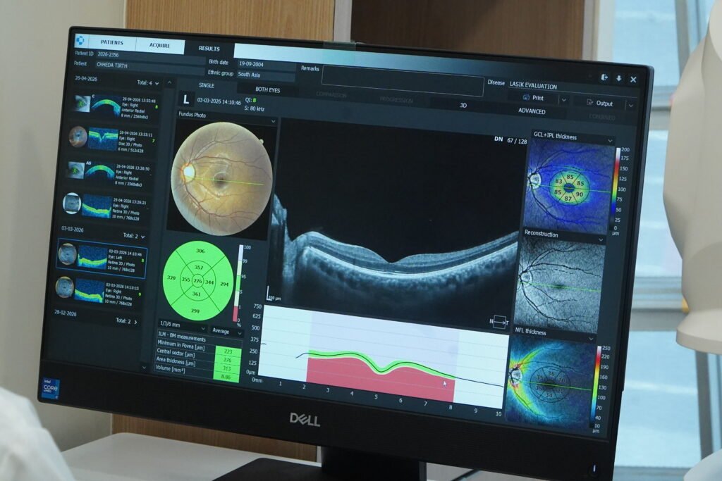
OCT — Optical Coherence Tomography — is the single most important imaging test in modern eye care. In simple words (OCT test kya hota hai?): it uses harmless light waves — no radiation, no injections, no touching the eye — to build a cross-sectional picture of your retina, the way a biopsy slide would, without cutting anything. Doctors call it an “optical biopsy.” Where a torch-light examination shows the surface of the retina, an OCT shows all ten microscopic layers — and disease almost always begins in the layers, not on the surface.
OCT in ophthalmology has done for the retina what MRI did for the brain. Before OCT (invented in 1991, clinical by the mid-1990s), retina specialists had to guess from surface appearance. Today, a macular OCT detects swelling of 20 micrometres — thinner than a human hair — and that early detection is exactly what saves vision in diabetic retinopathy, age-related macular degeneration (AMD) and glaucoma. If you have been searching for an optical coherence tomography test near me anywhere in Mumbai’s western suburbs — Andheri West, Lokhandwala, Versova, Juhu, Vile Parle, Oshiwara, Jogeshwari — this page tells you everything: the cost, the procedure, and the one question almost nobody asks: who reads your scan?
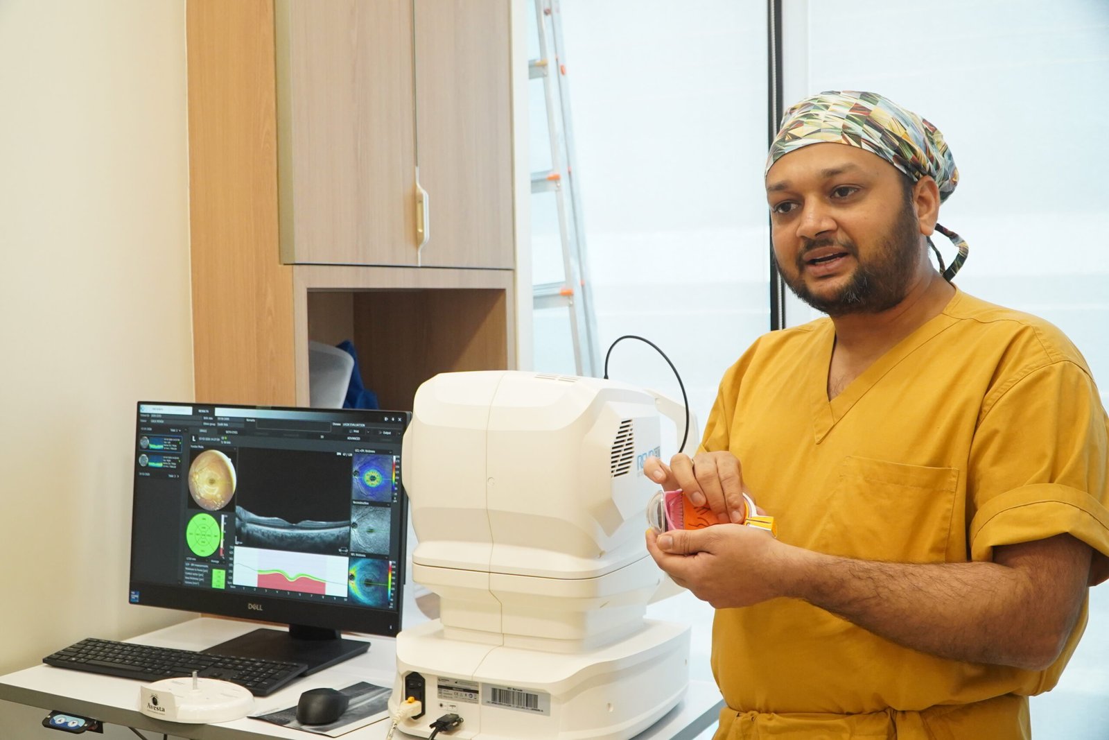
Dr. Jignesh Gala — retina specialist in Andheri West and FRCS (Glasgow)-trained vitreoretinal surgeon — uses OCT findings every single day to time injections, plan lasers, decide surgeries, and reassure patients whose scans are normal. His rule is simple: an OCT machine is only as good as the brain interpreting it. That is why at both his centres — Crystal Clear Eye Clinic and Topax Eye Hospital — scans are reported by the specialist himself, never outsourced to a technician.
2. Types of OCT Scans & What Each One Shows
“OCT” is a family of scans, not a single test. When a lab quotes you an eye OCT test price, ask which OCT they mean — the four scans below answer four different clinical questions:
| OCT Scan Type | What It Images | Main Use | Typical Mumbai Price (2026) |
|---|---|---|---|
| Macular OCT (retina OCT) | Cross-section of the central retina (macula) — all 10 layers + retinal thickness map | OCT test for macula: diabetic macular edema, AMD, CSR, macular hole, epiretinal membrane, vitreomacular traction | ₹1,500–3,000 per eye |
| RNFL / Optic Disc OCT | Retinal nerve fibre layer thickness around the optic nerve, compared against an age-matched normal database | OCT for glaucoma — detects nerve damage years before visual field loss | ₹2,000–3,500 both eyes |
| OCTA (OCT Angiography) | Retinal & choroidal blood flow — without any dye injection | Diabetic retinopathy vascular mapping, wet AMD choroidal neovascularization, vein occlusions | ₹3,500–5,000 per eye |
| Anterior Segment OCT | Cornea, iris, drainage angle, lens | Angle-closure glaucoma risk, corneal disease, premium IOL planning | ₹1,500–2,500 |
Most patients need only one or two of these. A diabetic coming for a retina checkup in Andheri typically needs a macular OCT; a glaucoma suspect needs RNFL analysis; a diabetic whose doctor wants to see the blood vessels without an injection needs OCTA. Beware of centres that sell every patient every scan — or worse, centres where the machine decides the “report” and no doctor ever looks at the raw images. More on that in Section 7.
3. OCT vs OCTA vs FFA vs Fundus Photo — Which Test Do You Need?
Patients often arrive with a prescription that says “fundus test” or “FFA” and ask whether an OCT is the same thing. It is not. These four tests answer different questions — and at a specialist retina clinic, they are combined, not substituted:
| Test | What It Shows | Dye/Injection? | Best For | Mumbai Price (2026) |
|---|---|---|---|---|
| OCT | Retinal layers in cross-section; thickness maps | No | Macular edema, AMD, holes, membranes, glaucoma RNFL | ₹1,500–3,500 |
| OCTA | Blood-flow map of retinal vessels without dye | No | Diabetic retinopathy vessels, wet AMD membranes, vein occlusion | ₹3,500–5,000 |
| FFA (Fundus Fluorescein Angiography) | Dye-based video of retinal circulation + leakage | Yes — IV dye in arm | Laser planning, vasculitis, complex vascular leakage | ₹3,000–6,000 |
| Fundus photography | Colour surface photo of the retina | No | Documentation, screening camps, baseline records — a fundus test for eyes in Mumbai usually means this | ₹500–1,500 |
Dr. Gala’s take: OCTA has replaced FFA in most follow-up situations — no injection, no nausea, no allergic risk, and repeatable at every visit. FFA still matters when planning laser for proliferative diabetic retinopathy or investigating inflammatory eye disease. If a centre pushes FFA on you without telling you why OCTA won’t do, ask questions.
4. 10 Retina & Eye Conditions an OCT Detects Early
An OCT is not a “general eye test” — it is a targeted diagnostic weapon. From OCT for diabetic retinopathy to glaucoma — here is what it catches, with the research Dr. Gala follows for each (full evidence library below):
- Diabetic macular edema (DME) — the #1 reason diabetics lose reading vision. OCT measures edema thickness to the micrometre and picks up fluid before vision drops; OCT biomarkers now predict which patients respond to which injection (PubMed evidence). This is the core OCT test for diabetic retinopathy (see our diabetic retinopathy guide for Andheri) and OCT test for macular edema.
- Diabetic retinopathy vascular damage — OCTA maps capillary dropout and microaneurysms without dye (evidence), which is why diabetic eye screening Mumbai (diabetic eye screening in Mumbai) at a retina centre should include OCTA, not just a fundus photo.
- Glaucoma — RNFL thinning on OCT appears years before visual field defects (evidence). An OCT test for glaucoma is the earliest warning system available; serial RNFL analysis tracks whether your treatment is actually working.
- Age-related macular degeneration (AMD) — drusen volume, subretinal fluid, and atrophy are all OCT biomarkers (evidence). The OCT test for macular degeneration / OCT test for AMD decides when ARMD treatment injections are needed and when they can safely be paused.
- Central serous chorioretinopathy (CSR/CSCR) — fluid under the retina, common in stressed professionals and steroid users; the OCT test for CSR confirms it in minutes and tracks resolution (evidence).
- Macular hole — OCT grades the hole (stage 1–4) and predicts surgical success (evidence). The OCT test for macular hole is mandatory before any vitrectomy.
- Epiretinal membrane (ERM) — the “cellophane wrinkle” on the macula; OCT shows whether it is just watching-worthy or vision-threatening (evidence). Search OCT test for epiretinal membrane and this is the scan you will get.
- Vitreomacular traction (VMT) — the jelly pulling on the retina; OCT shows whether it will release on its own or needs surgery (evidence).
- Retinal vein occlusion (BRVO/CRVO) — OCT quantifies macular swelling and monitors injection response at every visit.
- Choroidal diseases & high myopia — swept-source / enhanced-depth OCT images the choroid beneath the retina (evidence) — important in pachychoroid disease and myopic degeneration.
The research-aware difference: AI can now flag abnormal OCTs (AI-in-OCT evidence) — and Dr. Gala welcomes it as a second reader. But AI cannot examine you. He is fully aware of the latest research — unlike old practitioners who stopped reading journals years ago — and applies 2026’s biomarker evidence to every report he signs.
5. Who Needs an OCT & How Often
| You Are… | Recommended OCT | How Often |
|---|---|---|
| A diabetic (any duration, any sugar control) | Macular OCT ± OCTA — the real sugar ke patient ke liye OCT test | Once a year minimum; every 3–6 months if retinopathy exists; every 4–8 weeks during injection treatment |
| 40+ with family history of glaucoma / high eye pressure | RNFL + optic disc OCT (glaucoma test OCT near me — this is it) | Every 6–12 months |
| 50+, especially smoker or with family AMD history | Macular OCT (OCT test for AMD) | Yearly; immediately if distortion/blur appears |
| On long-term steroids, or stressed with sudden central blur | Macular OCT for CSR | At symptoms; then per recovery |
| High myope (−6 or more) | Macular OCT + peripheral retina check | Yearly |
| Anyone with sudden vision drop, distortion, or a curtain/shadow | Urgent macular OCT + dilated retina exam | Same day — do not wait |
If you are simply looking for an eye checkup near me after 40 — do it properly. A torch-and-chart “checkup” misses what an OCT catches. One baseline macular OCT + RNFL scan creates a permanent record against which every future change is measured. Telemedicine studies confirm OCT-based screening catches disease that routine camps miss (screening evidence).
6. The 5-Minute Procedure — Step by Step (OCT test kaise hota hai)
Patients ask OCT test mein kya hota hai (also searched as aankhon ka OCT test) — is there pain, injection, light flashes? Here is the honest walkthrough:
- Arrival (2 min): No fasting, no special preparation. Bring your glasses list and diabetes/BP medicines list.
- Dilating drops (only if needed, 15–20 min): For most macular OCTs, drops widen the pupil for a cleaner scan. RNFL scans often need no dilation at all. Near vision stays blurry for 2–3 hours afterwards — avoid driving yourself.
- The scan (30–60 seconds per eye): Chin on the rest, forehead against the bar, look at a small light. The machine hums and scans. No touch, no pain, no radiation, no dye. OCT test ke nuksan (harms)? Essentially none — it is just light. It is safe in pregnancy and safe repeatedly, which is why we can scan diabetics every month if needed.
- Instant images: Cross-sections and thickness maps appear immediately. OCT scan report normal ranges are built into the machine’s age-matched comparison database, and every OCT report explanation is done personally by Dr. Gala — he shows you your layers on the screen, in your language.
- Plan: Normal → reassurance + next screening date. Abnormal → treatment options explained the same visit (injection, laser, observation, or referral within his own hospitals if surgery is needed).
OCT test kaise karwaye Mumbai? Simply call 077188 85245 — same-day appointments are usually available at both Andheri West and Vile Parle East. Report in hand within the same visit.
7. Who Reads Your Scan? The Difference That Changes Diagnoses
The question that separates centres: At most diagnostic labs and aggregator-listed “eye test” centres in Mumbai, a technician operates the machine and the printed auto-report goes into your file. Some centres email scans to an outside doctor who has never seen you. At Crystal Clear Eye Clinic and Topax Eye Hospital, every OCT is acquired under and read by Dr. Jignesh Gala — a vitreoretinal surgeon who operates on the very diseases the scan detects.
Why it matters: an OCT report is not a blood test with a “normal range.” A small epiretinal membrane can be safely watched for years — or may be the reason your glasses never feel right. A diabetic macula can look “dry” on the report yet hide ischemia only a specialist suspects on the raw b-scans. Subretinal fluid in CSR looks similar to fluid in wet AMD to an algorithm — but the treatment is opposite. The scan is data; the diagnosis is a decision. Get the decision from the surgeon who will stand behind it.
8. OCT Test Cost in Mumbai & Andheri West (2026 Price Table)
Transparent, date-stamped (23 July 2026) — because patients deserve real numbers, not “call for price”:
| Scan | Typical Mumbai Range | Aggregator/Corporate Chains | At Dr. Gala’s Centres |
|---|---|---|---|
| Macular OCT (per eye) | ₹1,500–3,000 | ₹2,000–3,500 | Within range — reported by the retina specialist himself |
| RNFL / glaucoma OCT (both eyes) | ₹2,000–3,500 | ₹2,500–4,000 | Within range + specialist correlation with eye pressure & fields |
| OCTA (per eye) | ₹3,500–5,000 | ₹4,000–6,000 | Within range — dye-free vascular imaging |
| Anterior segment OCT | ₹1,500–2,500 | ₹2,000–3,000 | Within range |
| Fundus photography | ₹500–1,500 | ₹800–2,000 | Done when documentation adds value — not as a paid add-on by default |
Honest guidance: the OCT scan cost in Mumbai varies more by who interprets than by machine brand. A ₹1,200 scan misread by nobody-in-particular is the most expensive scan you will ever buy. The OCT eye test cost in Vile Parle or Andheri at Dr. Gala’s centres sits inside the normal city range — the difference is the vitreoretinal surgeon’s brain included in the price. Looking for an affordable OCT scan near me? Call 077188 85245 and ask for today’s exact price — you will get a number, not a runaround.
9. Insurance, Cashless & Reimbursement
A standalone diagnostic OCT on an OPD basis is usually not covered under standard Indian mediclaim OPD exclusions — that is true everywhere in Mumbai, not just here. However:
- When OCT is part of a procedure (pre-operative for surgery, or bundled with an intravitreal injection / laser that is covered), it is typically included in the package — and here Dr. Gala’s dual setup helps: direct cashless is done through Topax Eye Care (Vile Parle East), and third-party/TPA cashless is done through Crystal Clear Eye Clinic (Andheri West).
- OPD-cover policies & corporate health cards increasingly reimburse diagnostics — bring your card; the clinic provides proper GST invoices, machine-printed reports and image copies for smooth reimbursement claims.
- CGHS/reimbursement patients: rates are structured to stay within standard reference ranges; documentation is provided in the format claim offices expect.
10. Why Patients Choose Dr. Jignesh Gala for Their OCT

Dr. Jignesh Gala is a vitreoretinal and cataract surgeon — an FRCS (Glasgow)-trained retina specialist who has devoted his career to the back of the eye. He is the Founder & Director of both Crystal Clear Eye Clinic (Andheri West) and Topax Eye Hospital (Vile Parle East) — his own clinics, his own machines, his own name on every report.
✅ The best retina specialist in Andheri West reads your scan personally — not a technician, not an emailed-out report. A vitreoretinal specialist near me who also performs the injections, lasers and surgeries your scan may point to — continuity from image to treatment under one roof.
✅ Fully aware of the latest research — unlike old practitioners who stopped reading journals years ago. From OCT biomarkers that predict injection response to AI-assisted interpretation and swept-source choroidal imaging (110+ verified references below), he practises 2026’s retina care, not 2006’s.
✅ Both centres run current-generation OCT technology — patients searching for the best OCT machine in Mumbai get fast, high-definition scans with OCTA capability, compared visit-to-visit on the same machine for true progression tracking.
✅ Reports explained in your language — Hindi, Marathi, Gujarati or English — with the images on screen, so you leave understanding your own eye.
✅ Honest scanning policy: you get the scan you clinically need — not a bundle of paid add-ons. Sometimes the most valuable sentence in medicine is “your scan is normal; come back in a year.”
11. Locations — OCT Test in Andheri West & Vile Parle East

1) Crystal Clear Eye Clinic — Andheri West
📍 Address: Laram Centre CHS, A1-202, Swami Vivekanand Road, above Sunil Jewellers, near NADCO Shopping Centre, Railway Colony, Andheri West, Mumbai 400058
📞 Phone: 077188 85245 • 🕘 Open daily, closes 9 pm
💳 Third-party / TPA cashless is done through Crystal Clear Eye Clinic.
The answer to “Andheri West mein OCT test kahan karwaye?” / “aankh ka OCT test Andheri West mein kahan karwaye?” — minutes from Andheri station, Lokhandwala, Versova, Juhu, Oshiwara, DN Nagar, Seven Bungalows, Azad Nagar, Veera Desai Road, Jogeshwari West, Vile Parle West and Amboli. Patients from Bandra, Khar, Santacruz, Goregaon, Malad and Kandivali reach us in 20–30 minutes via Link Road or the metro. Searching OCT test in Andheri West or retina specialist in Andheri West? This is your stop.
2) Topax Eye Hospital — Vile Parle East
📍 Vile Parle East, Mumbai (full address on drjigneshgalaeye.com) • 📞 077188 85245
💳 Direct cashless is done through Topax Eye Care.
For aankh ka OCT test Vile Parle East, the OCT eye test cost in Vile Parle stays within the transparent ranges above. Convenient for Vile Parle East & West, Santacruz East, Khar, Bandra East, Kalina, Vakola, Juhu, and the airport corridor. As a full hospital, Topax handles everything a scan may lead to — injections, lasers, vitrectomy surgery — under Dr. Gala himself.
Distance is not a barrier: patients travel from Thane, Navi Mumbai, Borivali, Dadar and beyond for a specialist-read OCT. An OCT test for retina in Mumbai is only as valuable as the specialist interpreting it — and that is worth a short drive.
12. हिंदी • मराठी • ગુજરાતी — Patient Summaries
🇮🇳 हिंदी सारांश — आँखों का ओसीटी टेस्ट
ओसीटी टेस्ट क्या है? यह आँख की परदा (रेटिना) की परत-दर-परत तस्वीर लेने वाला एक बिल्कुल बेदर्द, 5 मिनट का स्कैन है — बिना इंजेक्शन, बिना रेडिएशन। ओसीटी टेस्ट के फायदे: डायबेटिक रेटिनोपैथी, ग्लूकोमा, AMD (उम्र से जुड़ी मैक्युलर बीमारी), मैक्युलर होल और रेटिना की सूजन जैसी बीमारियाँ शुरुआत में ही पकड़ी जाती हैं — जब इलाज आसान और असरदार होता है। मुंबई में ओसीटी टेस्ट की कीमत ₹1,500–3,500 तक होती है। रेटिना विशेषज्ञ अंधेरी वेस्ट में — डॉ. जिग्नेश गाला खुद आपकी रिपोर्ट पढ़ते हैं। मुंबई में डायबेटिक रेटिनोपैथी की जांच सहित हर स्कैन की रिपोर्ट उसी दिन, आपकी भाषा में समझाई जाती है। अपॉइंटमेंट: 077188 85245।
🚩 मराठी सारांश — ओसीटी टेस्ट
ओसीटी टेस्ट म्हणजे काय? डोळ्यांच्या पारदर्शक पडद्याचा (रेटिना) स्तर-स्तर स्कॅन — पूर्ण वेदनारहित, ५ मिनिटांचा, कोणताही इंजेक्शन किंवा रेडिएशन नाही. डायबेटिक रेटिनोपॅथी (मुंबईत डायबेटिक रेटिना तपासणी साठी), ग्लुकोमा, वयानुसार मॅक्युलर डिजेनरेशन (AMD), मॅक्युलर होल व रेटिनाच्या सूजेवरील लवकर निदान होते. मुंबईत ओसीटी ची किंमत ₹१,५००–३,५००. रेटिना विशेषज्ञ अंधेरी वेस्ट येथे — डॉ. जिग्नेश गाला स्वतः तुमचा ओसीटी अहवाल वाचतात व मराठीत समजावतात. भेटीसाठी: 077188 85245.
🟡 ગુજરાતી સારાંશ — ઓસીટી ટેસ્ટ
ઓસીટી ટેસ્ટ શું છે? આંખની રેટિનાનો સ્તર-દર-સ્તર સ્કેન — સંપૂર્ણ પીડારહિત, ૫ મિનિટનો, કોઈ ઈન્જેક્શન કે રેડિએશન વગર. ડાયાબેટિક રેટિનોપેથી, ગ્લૂકોમા, AMD, મેક્યુલર હોલ અને રેટિનાની સોજો વહેલા તબક્કે પકડાય છે. મુંબઈમાં ઓસીટીની કિંમત ₹૧,૫૦૦–૩,૫૦૦. રેટિના સ્પેશિયાલિસ્ટ અંધેરી વેસ્ટમાં — ડૉ. જિગ્નેશ ગાલા જાતે તમારો રિપોર્ટ વાંચીને ગુજરાતીમાં સમજાવે છે. એપોઈન્ટમેન્ટ: 077188 85245.
13. ❓ 12 Frequently Asked Questions (English + Hinglish)
1. OCT test dard hota hai kya? (Is the OCT eye test painful?)
Bilkul nahi. The machine never touches your eye — no injection, no dye, no radiation, no bright flash. You simply rest your chin and look at a small light for 30–60 seconds per eye. If dilating drops are used, they sting for a few seconds and blur near vision for 2–3 hours — that is the only discomfort. OCT test ke nuksan (harms)? Essentially none; it is just light.
2. OCT eye test kitne minute lagte hain? (How many minutes does it take?)
The scan itself takes 30–60 seconds per eye. With registration and (if needed) 15–20 minutes of dilation, plan for a 20–40 minute visit. Report in hand the same visit at both our centres.
3. OCT eye test cost in Andheri West – kitna kharcha aata hai?
Macular OCT ₹1,500–3,000 per eye; RNFL/glaucoma OCT ₹2,000–3,500 (both eyes); OCTA ₹3,500–5,000; anterior segment OCT ₹1,500–2,500 (prices verified 23 July 2026). At Crystal Clear Eye Clinic you pay within the normal city range — and the report is read by the retina specialist himself. OCT scan ki kimat confirm karne ke liye call 077188 85245 — today’s exact price, no runaround.
4. What does an OCT scan actually show?
Cross-sectional images of all ten retinal layers at micrometre resolution — fluid, swelling, holes, membranes, drusen, nerve fibre thickness — plus quantitative thickness maps compared against an age-matched normal database. It finds disease before symptoms, which is exactly when treatment works best.
5. OCT kyun zaroori hai? (Why is OCT necessary?)
Kyunke aankh ki andar ki bimari (diabetic retinopathy, glaucoma, AMD) pehle layers mein shuru hoti hai — surface par dikhti nahi. Torch-light check-up mein ye miss ho jati hai. OCT test ke fayde (benefits) ye hain: OCT detects swelling of 20 micrometres — thinner than a hair — months or years before vision drops. Early detection = simpler, cheaper, more successful treatment.
6. OCTA aur FFA mein kya difference hai?
Both image retinal blood vessels. OCTA does it with light alone — no injection, no allergy risk, repeatable every visit. FFA needs a dye injection in the arm (nausea/allergy possible) but shows live leakage and is still needed for laser planning in some cases. For most follow-ups, OCTA has replaced FFA.
7. Do I need dilatation for an OCT? Report kab milta hai?
Usually yes for macular scans (15–20 min for drops to work); RNFL/glaucoma scans often need no dilation. Report turant milta hai — images appear instantly, and Dr. Gala explains them on screen the same visit, in Hindi, Marathi, Gujarati or English.
8. How often should a diabetic get an OCT done?
Minimum once a year even with perfect sugar control. If diabetic retinopathy or macular edema exists: every 3–6 months. During anti-VEGF injection treatment: every 4–8 weeks — the scan, not the calendar, decides the next injection. Sugar ke patient ke liye OCT test — yaani retina ka OCT test — is not optional; it is the test that protects reading vision.
9. Can OCT detect glaucoma before vision is affected?
Yes — this is one of its biggest wins. RNFL thinning on OCT appears years before visual field defects develop. Serial scans then show whether treatment is truly stopping progression. After 40 — especially with family history or high eye pressure — a baseline RNFL OCT is strongly recommended.
10. OCT scan kahan karayein Andheri West mein?
Crystal Clear Eye Clinic — Laram Centre CHS, A1-202, S.V. Road, above Sunil Jewellers, near NADCO Shopping Centre, Andheri West 400058. Or at Topax Eye Hospital, Vile Parle East. Same phone for both: 077188 85245. Open daily, closes 9 pm. Same-day appointments usually available.
11. Is OCT safe for children, pregnant women and elderly patients?
Completely safe. No radiation (unlike CT scans), no dye, no contact. Children old enough to sit still for a minute can be scanned; it is safe in pregnancy; and it can be repeated as often as medically needed — which is why we can monitor treatment monthly without any risk accumulation.
12. OCT report mein RNFL thinning ya intraretinal fluid likha hai – matlab kya?
RNFL thinning = the nerve fibre layer around the optic nerve is thinner than expected for your age — an early glaucoma warning that needs pressure checks, fields and follow-up scans. Intraretinal fluid = swelling inside the retinal layers — commonly from diabetes, vein occlusion or AMD, usually treated with injections. Don’t panic over report words — bring the report to a retina specialist. OCT report samajhna (understanding the report) is a consultation, not a Google search — call 077188 85245 and Dr. Gala will explain exactly what yours means.
📖 Related Reading by Dr. Jignesh Gala
Keep learning from the same specialist who reads your scan:
- Intravitreal Injection in Andheri West, Mumbai — the complete guide
- Anti-VEGF Injection Cost in Mumbai — 2026 honest price guide
- Retina Laser Treatment in Andheri West / Mumbai
- Best Retina Specialist in Mumbai — Dr. Jignesh Gala
- Best Eye Specialist in Andheri, Mumbai
- Retina Treatment in Andheri
- Retina Treatment in Mumbai — Advanced Retinal Care
- Retina Surgery in Mumbai — Your Complete Guide to Saving Your Sight
- Cashless Eye Surgery in Mumbai
- Surgical Videos by Dr. Jignesh Gala
- Inside My Six Years as a BMJ Case Reports Peer Reviewer
- Diabetic Retinopathy in Andheri
- Diabetic Eye Care in Mumbai
- Diabetic Macular Edema (DME) Treatment in Mumbai
- Macular Edema Treatment in Andheri West
- ARMD Treatment in Andheri West, Mumbai
- Vitreoretinal Treatment in Andheri
- Best Retina Specialist in Mumbai
- Best Vitrectomy Surgery in Mumbai — Expert Retinal Care
- Cashless Eye Surgery in Mumbai
- Sudden Floaters? Retina Check-up in Mumbai
- What Is an Intravitreal Eye Injection? Explained by Dr. Jignesh Gala
Your retina can’t wait. Neither should you.
Whether you searched for an affordable OCT scan near me, an affordable retina OCT scan in Mumbai, a glaucoma test OCT near me, an OCT scan for diabetic retinopathy Mumbai, or checked the OCT test cost in Mumbai — you have found transparent pricing and specialist-read scans. One 5-minute scan today can protect decades of reading vision.
📍 Andheri West: Laram Centre CHS, A1-202, S.V. Road, above Sunil Jewellers, near NADCO Shopping Centre, Mumbai 400058
📍 Vile Parle East: Topax Eye Hospital • Open daily, closes 9 pm
Dr. Jignesh Gala, FRCS (Glasgow) — Vitreoretinal & Cataract Surgeon • drjigneshgalaeye.com • crystalcleareye.in
Medical disclaimer: This page is educational and does not replace a personal consultation. Prices are indicative, verified on 23 July 2026, and may change; please confirm at booking. Every eye is different — treatment decisions are made only after examination by Dr. Jignesh Gala.
14. Research & Evidence Library — 110+ Verified References
Dr. Gala reads the research behind every scan he interprets. Every link below is verified live (July 2026) — click through to read the same evidence he follows.
🔬 OCT (Optical Coherence Tomography) Clinical Evidence — PubMed (50 verified)
Diabetic Macular Edema Biomarkers
- Multimodal imaging in diabetic retinopathy and macular edema: An update about biomarkers — Survey of ophthalmology, 2024
- Persistent diabetic macular edema: Definition, incidence, biomarkers, and treatment methods — Survey of ophthalmology, 2023
- Baseline OCT Biomarkers Associated with Visual Acuity in Diabetic Macular Edema: A Systematic Review and Meta-analysis — Ophthalmology, 2026
- Optical coherence tomography in the management of diabetic macular oedema — Progress in retinal and eye research, 2024
- Imaging and Biomarkers in Diabetic Macular Edema and Diabetic Retinopathy — Current diabetes reports, 2019
Oct Angiography In Diabetic Retinopathy
- OCT angiography 2023 update: focus on diabetic retinopathy — Acta diabetologica, 2024
- New insights into diabetic retinopathy by OCT angiography — Diabetes research and clinical practice, 2018
- Optical coherence tomography angiography in diabetic retinopathy: an updated review — Eye (London, England), 2021
- Association of OCT Angiography-Detected Intraretinal Microvascular Abnormalities with Diabetic Retinopathy Severity — Ophthalmology. Retina, 2026
- COMPARATIVE ANALYSIS OF OCT AND OCT ANGIOGRAPHY CHARACTERISTICS IN EARLY DIABETIC RETINOPATHY — Retina (Philadelphia, Pa.), 2023
Glaucoma – Retinal Nerve Fiber Layer
- Retinal Nerve Fiber Layer Optical Texture Analysis: Involvement of the Papillomacular Bundle and Papillofoveal Bundle in Early Glaucoma — Ophthalmology, 2022
- Anatomical compensation of retinal nerve fiber layer improves the detection of glaucoma between ethnicities — Annals of the New York Academy of Sciences, 2024
- Racial Differences in Diagnostic Accuracy of Retinal Nerve Fiber Layer Thickness in Primary Open-Angle Glaucoma — American journal of ophthalmology, 2024
- Peripapillary Retinal Nerve Fiber Layer Vascular Microcirculation in Glaucoma Using Optical Coherence Tomography-Based Microangiography — Investigative ophthalmology & visual science, 2016
- Evaluating the retinal nerve fiber layer in glaucoma with scanning laser polarimetry — Archives of ophthalmology (Chicago, Ill. : 1960), 1999
Telemedicine / Screening For Diabetic Retinopathy
- Review of smartphone funduscopy for diabetic retinopathy screening — Survey of ophthalmology, 2024
- Comparison of Telemedicine Screening of Diabetic Retinopathy by Mydriatic Smartphone-Based vs Nonmydriatic Tabletop Camera-Based Fundus Imaging — Journal of vitreoretinal diseases, 2021
- The role of tele-ophthalmology in diabetic retinopathy screening — Journal of optometry, 2020
- Cost-effectiveness of a National Telemedicine Diabetic Retinopathy Screening Program in Singapore — Ophthalmology, 2016
- Feasibility of telemedicine in detecting diabetic retinopathy and age-related macular degeneration — Seminars in ophthalmology, 2015
Artificial Intelligence In Oct Interpretation
- [OCT biomarkers in diabetic maculopathy and artificial intelligence] — Die Ophthalmologie, 2024
- Artificial intelligence for retinal diseases — Asia-Pacific journal of ophthalmology (Philadelphia, Pa.), 2024
- Artificial Intelligence and Imaging Processing in Optical Coherence Tomography and Digital Images in Uveitis — Ocular immunology and inflammation, 2022
- Application of Artificial Intelligence in Targeting Retinal Diseases — Current drug targets, 2020
- A comparison of deep learning performance against health-care professionals in detecting diseases from medical imaging: a systematic review and meta-analysis — The Lancet. Digital health, 2019
Macular Hole & Epiretinal Membrane
- Epiretinal membrane: an overview and update — Japanese journal of ophthalmology, 2024
- In-Depth Analysis of Preoperative OCT Markers as Prognostic Factors for Lamellar Macular Holes and Epiretinal Membrane Foveoschisis — Ophthalmology. Retina, 2024
- Macular hole with epiretinal proliferation: diagnostic value of en-face optical coherence tomography and clinical characteristics — Graefe’s archive for clinical and experimental ophthalmology = Albrecht von Graefes Archiv fur klinische und experimentelle Ophthalmologie, 2024
- The clinical and pathogenic significance of atypical epiretinal tissue in macular hole — Graefe’s archive for clinical and experimental ophthalmology = Albrecht von Graefes Archiv fur klinische und experimentelle Ophthalmologie, 2022
- Optical coherence tomography in the preoperative and postoperative management of macular hole and epiretinal membrane — The British journal of ophthalmology, 2014
Central Serous Chorioretinopathy
- Central serous chorioretinopathy and the sclera: what we have learned so far — Japanese journal of ophthalmology, 2024
- Can we classify central serous chorioretinopathy better? Yes we can — Eye (London, England), 2022
- Diagnostic accuracy of OCT angiography for macular neovascularization in central serous chorioretinopathy: A systematic review and meta-analysis — Acta ophthalmologica, 2024
- Regression patterns of central serous chorioretinopathy using en face optical coherence tomography — Graefe’s archive for clinical and experimental ophthalmology = Albrecht von Graefes Archiv fur klinische und experimentelle Ophthalmologie, 2022
- Central Serous Chorioretinopathy Analyzed by Multimodal Imaging — Translational vision science & technology, 2021
Age-Related Macular Degeneration Biomarkers
- OCT Prognostic Biomarkers for Progression to Late Age-related Macular Degeneration: A Systematic Review and Meta-analysis — Ophthalmology. Retina, 2024
- Baseline OCT Biomarkers Predicting Visual Outcomes in Neovascular Age-Related Macular Degeneration: A Meta-Analysis — Ophthalmology, 2025
- Choroidal biomarkers in age-related macular degeneration — Survey of ophthalmology, 2025
- Practical guidance for imaging biomarkers in exudative age-related macular degeneration — Survey of ophthalmology, 2023
- Predicting Progression of Age-Related Macular Degeneration Using OCT and Fundus Photography — Ophthalmology. Retina, 2021
Vitreomacular Traction
- Vitreomacular traction – a review — Eye (London, England), 2025
- Vitreomacular traction in diabetic retinopathy — Japanese journal of ophthalmology, 2024
- Vitreomacular Traction Surgery from the DISCOVER Study: Intraoperative OCT Utility, Ellipsoid Zone Dynamics, and Outcomes — Ophthalmic surgery, lasers & imaging retina, 2021
- A Study of the Natural History of Vitreomacular Traction Syndrome by OCT — Ophthalmology, 2018
- The International Vitreomacular Traction Study Group classification of vitreomacular adhesion, traction, and macular hole — Ophthalmology, 2013
Swept-Source Oct / Enhanced Depth Imaging Choroid
- Choroidal biomarkers — Indian journal of ophthalmology, 2018
- Enhanced depth imaging in swept-source optical coherence tomography: Improving visibility of choroid and sclera, a masked study — European journal of ophthalmology, 2020
- Choroidal imaging biomarkers — Survey of ophthalmology, 2019
- Enhanced depth imaging-OCT of the choroid: a review of the current literature — Graefe’s archive for clinical and experimental ophthalmology = Albrecht von Graefes Archiv fur klinische und experimentelle Ophthalmologie, 2014
- Enhanced depth imaging optical coherence tomography — Ophthalmic surgery, lasers & imaging : the official journal of the International Society for Imaging in the Eye, 2011
🏛️ Global Authorities & Trial Registries (40 verified)
AAO (aao.org) — 7 links
- AAO: Anti-VEGF Treatments — Plain-language explainer of what anti-VEGF eye injections are, conditions treated, and what to expect during the shot.
- AAO: Diabetic Retinopathy: Causes, Symptoms, Treatment — Patient guide to diabetic eye disease stages, diagnosis (OCT), and treatments including anti-VEGF injections.
- AAO: Understanding Macular Degeneration — Patient overview of dry vs wet AMD, AREDS2 vitamins, anti-VEGF therapy, and Amsler grid self-monitoring.
- AAO: Retinal Vein Occlusion — Explains BRVO vs CRVO, macular edema, and anti-VEGF/laser treatment options for patients.
- AAO: Diabetic Retinopathy PPP 2024 — AAO's official evidence-based clinical guideline for diabetic retinopathy care (free PDF).
- AAO: Age-Related Macular Degeneration PPP 2024 (Updated 2026) — AAO's official AMD clinical guideline, including anti-VEGF treatment recommendations.
- AAO: Retinal Vein Occlusions PPP 2024 (Updated 2026) — AAO's official RVO clinical guideline covering anti-VEGF agents and aflibercept 8 mg.
EyeWiki (eyewiki.org) — 5 links
- EyeWiki: Intravitreal Injections — Physician-reviewed reference on injection technique, indications, and complications.
- EyeWiki: Aflibercept — Drug monograph on Eylea: mechanism, dosing, trial evidence.
- EyeWiki: Ranibizumab — Drug monograph on Lucentis: mechanism, indications, key trials.
- EyeWiki: Faricimab — Drug monograph on Vabysmo, the dual VEGF/Ang-2 inhibitor.
- EyeWiki: Endophthalmitis — Reference on the most serious injection complication: signs, management, prognosis.
ASRS (asrs.org) — 5 links
- ASRS: Retina Health Series: Intravitreal Injections (fact sheet 30, PDF) — Patient fact sheet on what happens during an eye injection and its risks.
- ASRS: Retina Health Series: Age-Related Macular Degeneration (fact sheet 16, PDF) — Patient fact sheet on AMD symptoms, testing, and anti-VEGF treatment regimens.
- ASRS: Retina Health Series: Macular Edema (fact sheet 13, PDF) — Patient fact sheet on macular edema causes (incl. diabetes/DME) and treatments.
- ASRS: Retina Health Series: Central Retinal Vein Occlusion (fact sheet 21, PDF) — Patient fact sheet on CRVO and its anti-VEGF/laser management.
- ASRS: Diabetic Retinopathy (patient disease page) — ASRS patient portal page on diabetic retinopathy symptoms, causes, and treatments.
Retinal Physician (retinalphysician.com) — 3 links
- Retinal Physician: Comparing Intravitreal Injection Protocols and Practices (Sept 2010) — Expert roundtable on injection technique nuances: anesthesia, asepsis, needle handling, post-op care.
- Retinal Physician: Antisepsis for Intravitreal Injections (April 2023) — How povidone-iodine antisepsis lowers endophthalmitis risk from injections.
- Retinal Physician: Endophthalmitis Then and Now (May 2025) — Historical-to-modern review of endophthalmitis prophylaxis and management.
Landmark journal article pages (DOI-resolving) — 6 links
- Ophthalmology (AAO journal): Heier JS et al. — Intravitreal Aflibercept (VEGF Trap-Eye) in Wet AMD (VIEW 1/VIEW 2), 2012 — Pivotal trials that established aflibercept 2 mg for wet AMD.
- Ophthalmology: Dugel PU et al. — HAWK and HARRIER: Phase 3 Trials of Brolucizumab for nAMD, 2020 — Trials leading to brolucizumab (Beovu) approval.
- Lancet: Heier JS et al. — Faricimab in nAMD (TENAYA and LUCERNE), 2022 — Showed faricimab durability up to 16-week intervals in wet AMD.
- Lancet: Wykoff CC et al. — Faricimab in DME (YOSEMITE and RHINE), 2022 — Pivotal faricimab trials in diabetic macular edema.
- Ophthalmology: CATT Research Group — Ranibizumab and Bevacizumab for nAMD: Two-Year Results, 2012 — Head-to-head comparison of Lucentis vs Avastin for wet AMD.
- Ophthalmology: Wells JA et al. (DRCR.net Protocol T) — Aflibercept, Bevacizumab, or Ranibizumab for DME, 2016 — Comparative-effectiveness trial guiding anti-VEGF choice in DME.
ClinicalTrials.gov (all confirmed via official v2 API) — 10 links
- ClinicalTrials.gov: YOSEMITE — Faricimab in DME (COMPLETED) — Phase 3 faricimab trial in diabetic macular edema.
- ClinicalTrials.gov: RHINE — Faricimab in DME (COMPLETED) — Sister phase 3 faricimab DME trial (official title: "A Study to Evaluate the Efficacy and Safety of Faricimab (RO6867461) in Participants With Diabetic Macular Edema").
- ClinicalTrials.gov: PHOTON — Aflibercept 8 mg in DME (COMPLETED) — Phase 3 high-dose aflibercept in diabetic eye disease.
- ClinicalTrials.gov: PULSAR — Aflibercept 8 mg in wet AMD (COMPLETED) — Phase 3 high-dose aflibercept in neovascular AMD.
- ClinicalTrials.gov: TENAYA — Faricimab in nAMD (COMPLETED) — Phase 3 faricimab in neovascular AMD.
- ClinicalTrials.gov: LUCERNE — Faricimab in nAMD (COMPLETED) — Sister phase 3 faricimab nAMD trial.
- ClinicalTrials.gov: HAWK — Brolucizumab (RTH258) vs aflibercept, Study 1 (COMPLETED) — Phase 3 brolucizumab trial in wet AMD.
- ClinicalTrials.gov: HARRIER — Brolucizumab (RTH258) vs aflibercept, Study 2 (COMPLETED) — Sister phase 3 brolucizumab trial.
- ClinicalTrials.gov: Archway — Port Delivery System (Susvimo) with ranibizumab, wet AMD (COMPLETED) — Phase 3 refillable ranibizumab implant vs monthly injections.
- ClinicalTrials.gov: VIEW 1 — VEGF Trap-Eye (aflibercept) in wet AMD (COMPLETED) — Pivotal aflibercept wet AMD trial.
NEI (nei.nih.gov) — 2 links
- NEI: Age-Related Macular Degeneration — NIH patient resource on AMD types, risk factors, and treatment.
- NEI: Diabetic Retinopathy — NIH patient resource on diabetic eye disease and injections/laser/surgery.
MedlinePlus — 2 links
- MedlinePlus: Macular Degeneration — Curated NIH patient information hub on AMD.
- MedlinePlus: Diabetic Eye Problems — Patient hub on diabetic retinopathy and related eye complications.
📚 Related Retina Evidence — PubMed
Faricimab (Vabysmo) Trials
- Efficacy, durability, and safety of intravitreal faricimab up to every 16 weeks for neovascular age-related macular degeneration (TENAYA and LUCERNE): two randomised, double-masked, phase 3, non-inferiority trials. Lancet (London, England), 2022.
- TENAYA and LUCERNE: Two-Year Results from the Phase 3 Neovascular Age-Related Macular Degeneration Trials of Faricimab with Treat-and-Extend Dosing in Year 2. Ophthalmology, 2024.
- Efficacy, durability, and safety of intravitreal faricimab with extended dosing up to every 16 weeks in patients with diabetic macular oedema (YOSEMITE and RHINE): two randomised, double-masked, phase 3 trials. Lancet (London, England), 2022.
- Four-Year Outcomes of Faricimab in Diabetic Macular Edema: Results from the RHONE-X Extension Trial. Ophthalmology, 2026.
- Simultaneous Inhibition of Angiopoietin-2 and Vascular Endothelial Growth Factor-A with Faricimab in Diabetic Macular Edema: BOULEVARD Phase 2 Randomized Trial. Ophthalmology, 2019.
- Efficacy and Safety of Faricimab for Macular Edema due to Retinal Vein Occlusion: 24-Week Results from the BALATON and COMINO Trials. Ophthalmology, 2024.
- Anatomic Outcomes with Faricimab vs Aflibercept in Head-to-Head Dosing Phase of the TENAYA/LUCERNE Trials in Neovascular Age-related Macular Degeneration. Ophthalmology, 2025.
- Faricimab Treat-and-Extend Dosing for Macular Edema Due to Retinal Vein Occlusion: 72-Week Results from the BALATON and COMINO Trials. Ophthalmology. Retina, 2025.
- Faricimab for neovascular age-related macular degeneration and diabetic macular edema: from preclinical studies to phase 3 outcomes. Graefe's archive for clinical and experimental ophthalmology = Albrecht von Graefes Archiv fur klinische und experimentelle Ophthalmologie, 2024.
Aflibercept 8 Mg (Photon / Pulsar)
- Aflibercept 8 mg versus Faricimab Treat-and-Extend for Diabetic Macular Edema or Neovascular Age-Related Macular Degeneration: A Bayesian Fixed-Effect Network Meta-analysis of Clinical Trials. Ophthalmology and therapy, 2025.
- A Matching-Adjusted Comparison of Faricimab and Aflibercept 8 mg in Neovascular Age-Related Macular Degeneration and Diabetic Macular Edema. Ophthalmic research, 2026.
- Economic Benefit of Aflibercept 8 mg Versus Faricimab for Neovascular Age-Related Macular Degeneration or Diabetic Macular Edema in the US. Ophthalmology and therapy, 2025.
- Intraocular Pressure Outcomes with Intravitreal Aflibercept 8 mg: A 96-Week Pooled Analysis from PULSAR and PHOTON. Ophthalmology and therapy, 2026.
- A Response to: Letter to the Editor Regarding "Aflibercept 8 mg Versus Faricimab Treat-and-Extend for Diabetic Macular Edema or Neovascular Age-Related Macular Degeneration: A Bayesian Fixed-Effect Network Meta-analysis of Clinical Trials". Ophthalmology and therapy, 2026.
- Effect of High-Dose Intravitreal Aflibercept, 8 mg, in Patients With Neovascular Age-Related Macular Degeneration: The Phase 2 CANDELA Randomized Clinical Trial. JAMA ophthalmology, 2023.
- High-Dose 8 mg Aflibercept for Neovascular Age-Related Macular Degeneration: Who Is Being Treated with This New Agent? Life (Basel, Switzerland), 2025.
- Aflibercept high-dose (8mg) related intraocular inflammation (IOI) – a case series. BMC ophthalmology, 2024.
- Clinical Outcomes of High-Dose Aflibercept 8 mg in Polypoidal Choroidal Vasculopathy: A Systematic Review and Meta-Analysis. Cureus, 2026.
- Initial Functional and Anatomical Outcomes of High-dose Aflibercept 8 mg in Exudative Neovascular Age-related Macular Degeneration. Ophthalmology. Retina, 2025.
Treat-And-Extend Anti-Vegf
- Treat-and-extend dosing of intravitreal anti-VEGF agents in neovascular age-related macular degeneration: a meta-analysis. Eye (London, England), 2023.
- TREAT-AND-EXTEND REGIMENS WITH ANTI-VEGF AGENTS IN RETINAL DISEASES: A Literature Review and Consensus Recommendations. Retina (Philadelphia, Pa.), 2015.
- Vitrectomy as an Adjunct to Treat-and-Extend Anti-VEGF Injections for Diabetic Macular Edema: The Vitrectomy in Diabetic Macular Oedema (VIDEO) Randomized Clinical Trial. JAMA ophthalmology, 2024.
- Efficacy, safety, and treatment burden of treat-and-extend versus alternative anti-VEGF regimens for nAMD: a systematic review and meta-analysis. Eye (London, England), 2023.
- Treat and Extend Treatment Interval Patterns with Anti-VEGF Therapy in nAMD Patients. Vision (Basel, Switzerland), 2019.
- A Systematic Review of the Treat and Extend Treatment Regimen with Anti-VEGF Agents for Neovascular Age-Related Macular Degeneration. Ophthalmology and therapy, 2017.
- The Role of Intravitreal Corticosteroids in the Treatment of DME: Predictive OCT Biomarkers. International journal of molecular sciences, 2022.
- Progress and Challenges of Anti-VEGF Agents and Their Sustained-Release Strategies for Retinal Angiogenesis. Drug design, development and therapy, 2022.
- Treat and Extend Anti-VEGF Treatment Regimens for Neovascular Age-Related Macular Degeneration: To Stop or Not to Stop? Clinical & experimental ophthalmology, 2025.
- Treat-and-extend regimens of anti-vascular endothelial growth factor therapy for retinal vein occlusions: a systematic review and meta-analysis. Acta ophthalmologica, 2022.
Intravitreal Injection Real-World India
- Real-World Evidence in the Management of Diabetic Macular Edema with Intravitreal Anti-VEGFs in Asia: A Systematic Literature Review. Clinical ophthalmology (Auckland, N.Z.), 2022.
- Real-World Safety Outcomes of Intravitreal Ranibizumab Biosimilar (Razumab) Therapy for Chorioretinal Diseases. Ophthalmology and therapy, 2021.
- REal-world treatment outcomes after delayed intRavitreal therapy in center-involving diabetic macular edema – RETORT study. International journal of retina and vitreous, 2023.
- "Real world outcomes of intravitreal brolucizumab for persistent diabetic macular edema". International journal of retina and vitreous, 2025.
- Real-world experience of brolucizumab in nAMD. Indian journal of ophthalmology, 2024.
- Real-world outcomes of intravitreal anti-vascular endothelial growth factor monotherapy in proliferative type 2 macular telangiectasia. Graefe's archive for clinical and experimental ophthalmology = Albrecht von Graefes Archiv fur klinische und experimentelle Ophthalmologie, 2021.
- Ranizurel safety evaluation in real-world -(RaSER) study. American journal of ophthalmology case reports, 2022.
- Initial experience with brolucizumab for neovascular age-related macular degeneration (nAMD) in India – Multicentric, real-world study. Indian journal of ophthalmology, 2024.
- Real-world evidence of safety profile of intravitreal bevacizumab (Avastin) in an Indian scenario. Indian journal of ophthalmology, 2017.
- Factors determining compliance with recommended intravitreal injections for retinal diseases among out-of-pocket, fully subsidized (non-paying), and insured patients: Real-world evidence from India. Indian journal of ophthalmology, 2025.